. 2002 Sep;13(9):3281-93.
doi: 10.1091/mbc.e02-01-0005.

Calcineurin, a calcium/calmodulin-dependent protein phosphatase, is involved in movement, fertility, egg laying, and growth in Caenorhabditis elegans

Affiliations

Calcineurin, a calcium/calmodulin-dependent protein phosphatase, is involved in movement, fertility, egg laying, and growth in Caenorhabditis elegans

Jaya Bandyopadhyay et al. Mol Biol Cell. 2002 Sep.

Abstract

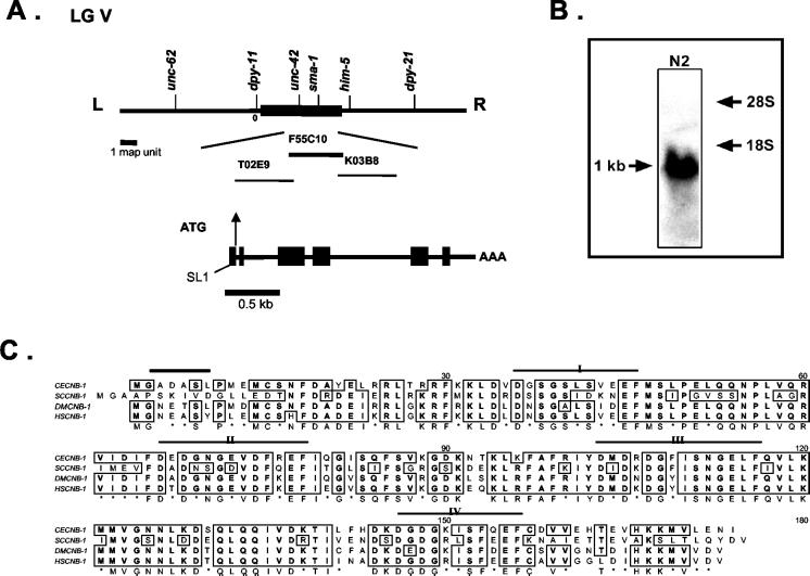
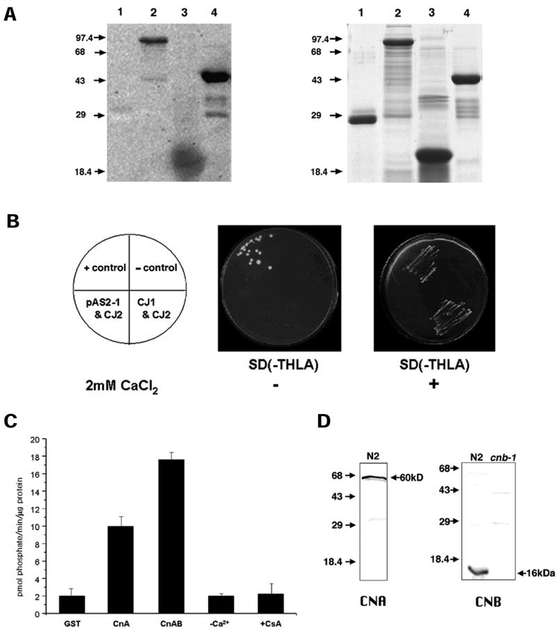
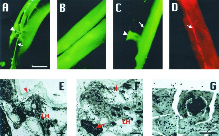
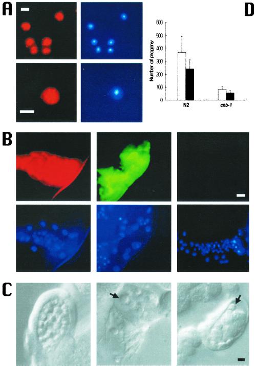
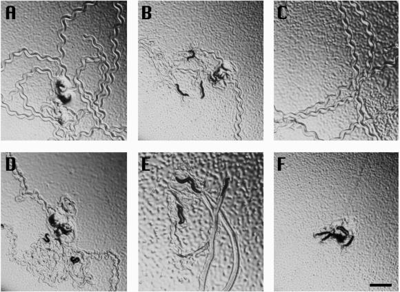
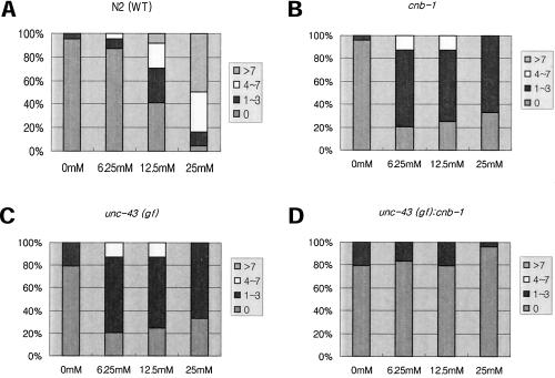
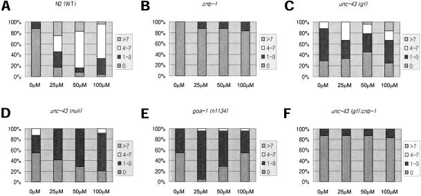
Calcineurin is a Ca(2+)-calmodulin-dependent serine/threonine protein phosphatase that has been implicated in various signaling pathways. Here we report the identification and characterization of calcineurin genes in Caenorhabditis elegans (cna-1 and cnb-1), which share high homology with Drosophila and mammalian calcineurin genes. C. elegans calcineurin binds calcium and functions as a heterodimeric protein phosphatase establishing its biochemical conservation in the nematode. Calcineurin is expressed in hypodermal seam cells, body-wall muscle, vulva muscle, neuronal cells, and in sperm and the spermatheca. cnb-1 mutants showed pleiotropic defects including lethargic movement and delayed egg-laying. Interestingly, these characteristic defects resembled phenotypes observed in gain-of-function mutants of unc-43/Ca(2+)-calmodulin-dependent protein kinase II (CaMKII) and goa-1/G(o)-protein alpha-subunit. Double mutants of cnb-1 and unc-43(gf) displayed an apparent synergistic severity of movement and egg-laying defects, suggesting that calcineurin may have an antagonistic role in CaMKII-regulated phosphorylation signaling pathways in C. elegans.


Figures

Figure 1
Figure 1
Genomic organization of calcineurin genes in C. elegans. (A) Genetic and physical maps of the cnb-1 region. The cnb-1 locus relative to the nearby genetic markers on the gene cluster region of LG V is shown. cnb-1, which is mapped to the cosmid F55C10 (Accession No. Z74036), and the neighboring cosmids are indicated. The predicted F55C10.1 gene (cnb-1) structure has been revised and is presently shown to contain six exons (black boxes). (B) Northern blot analysis detects a 1-kb band in RNA samples of wild-type N2 worms. (C) Amino acid sequence alignment for calcineurin B of C. elegans (CECNB-1), Drosophila (DMCNB-1, Accession No. P48451), human (HSCNB-1, Accession No. NP000936), and yeast (SCCNB-1, Accession No. NP_012731). Regions of identity among calcineurin B homologues are boxed. Four Ca2+-binding sites are shown (I–IV). The myristoylation signature is indicated by a black bar.
Figure 2
Figure 2
Biochemical characterization of C. elegans calcineurin. (A) cnb-1 is a Ca2+-binding protein. Ca2+-binding assay was performed with GST (glutathione-S-transferase) (lane 1), C. elegans calsequestrin, GST-CSQ-1 (lane 2), bovine CaM (lane 3), and GST-CNB-1 (lane 4). 45Ca2+-labeled proteins are shown by the dark signals on the x-ray film. GST (lane 1) is used as a negative control. A corresponding gel shown in the right panel is stained with Coomassie Blue. Molecular weight markers (in kDa) are denoted by arrows. (B) Interaction between CNA-1 and CNB-1 in the yeast two-hybrid system. Growth of yeast transformants on SD −Trp −His −Leu −Ade with 5 mM 3-AT (panel 2) and 5 mM 3-AT + 2 mM CaCl2 (panel 3) are shown. Plates supplemented with 5 mM 3-AT + 2 mM CaCl2 select for a positive interaction between the fusion proteins. Positive interaction between AD/T-antigen and DNA-BD/p53 fusion proteins and the negative or no interaction between AD/T-antigen and DNA-BD/lamin C were used as positive (+ control), and negative controls (− control), respectively. The array of the yeasts containing the different constructs is indicated in the schemes shown in panel 1. (C) Serine/threonine phosphatase activity of CNA-1. Dephosphorylation of the phosphopeptide by GST, GST-CNA-1 (CnA), and GST-CNA-1 + GST-CNB-1 (CnAB) are shown. Ca2+-dependence of the phosphatase activity (−Ca2+) was determined by adding 0.2 mM EGTA (Ca2+-chelator) during the CnAB reaction. Inhibition of phosphatase activity was determined by adding 1 μM each of CsA and bovine cyclophilin (+CsA) in the CnAB reaction mixture. Assays were performed in triplicate with equimolar concentrations of CNA-1 and CNB-1. (D) Western blot analysis with anti–CNA-1 and anti–CNB-1 antibodies. Worm protein extracts prepared from N2 wild-type worms or cnb-1(jh103) mutants (CNB, lane 2). The band is absent in the cnb-1(jh103) deletion worms indicating they are functionally null mutants.
Figure 3
Figure 3
Expression and localization of calcineurin. Images show only representative expression. CNA-1::GFP expressed in (A) ventral nerve cord (arrow) and vulva muscles (arrowhead) and (B) body-wall muscles of the midbody region. CNB-1::GFP expresses in spermatheca (C; arrowhead) and intestine (C; arrow). Nerve cord expression can also be observed in this image. (D) Immunostaining of wild-type worms with anti–CNB-1 antibodies detected CNB-1 in hypodermal seam cells (arrow). Bar, 20 μm. (EG) Immunogold electron micrograph (EM) showing the subcellular localization of CNB-1. (E) Transverse section through the midbody of a hermaphrodite showing CNB-1 localization as indicated by the signals of gold particles in the seam cells of the lateral hypodermis (LH) closely apposed to the cuticle (arrowhead). (F) Another region of seam cell showing signals. Lateral hypodermis (LH), cuticle (arrowhead), and excretory canal (arrow) are labeled. (G) Localization of CNB-1 in a wild-type male gonad. Distinct and scattered signals of CNB-1 are observed mainly in the cytoplasmic regions of the cellularized spermatids. Bar, 1 μm.
Figure 4
Figure 4
Calcineurin in sperm and the spermatheca. (A) Immunostaining of wild-type sperm with anti-CNA-1 (top left panel) and anti–CNB-1 (bottom left panel) antibodies and the respective nuclei are shown by DAPI staining (right panels). Bar, 2 μm. (B) Immunostaining of wild-type and cnb-1 mutant spermatheca with calcineurin antibodies. Anti-CNA-1 (top left panel) and anti–CNB-1 (top middle panel) antibodies stain wild-type spermatheca. Spermatheca of cnb-1 mutant worms do not show any anti–CNB-1 staining (top right panel). DAPI staining of spermatheca is shown in the bottom panels. Bar, 20 μm. (C) Sperm and spermatheca defects. Compared with wild-type (left panel) cnb-1 mutant worms display defects in sperm and the spermatheca. cnb-1 sperm (middle and right panel). Arrows point to cnb-1 sperm, which are fewer in number and show a smoother appearance compared with N2 sperm (left panel). The surrounding cnb-1 spermatheca appears to contain debris. (D) Mating of N2 and cnb-1 hermaphrodites with N2 males. Three N2 males were mated with one N2 or one cnb-1 hermaphrodite for 2 days. Total progeny and outcrossed progeny are indicated by white and black bars, respectively.
Figure 5
Figure 5
Locomotion defects in mutants of cnb-1 and unc-43. Five animals were placed in the center of a bacterial lawn and photographed 5 min later. (A) Tracks of movement by wild-type animals; (B) cnb-1 mutants; (C) rescued cnb-1 mutants; (D) unc-43(lf) mutants; (E) unc-43(gf) mutants; (F) cnb-1; unc-43(gf) double mutants. Bar, 1 mm.
Figure 6
Figure 6
Dose-dependent serotonin-mediated egg laying. Color-coded area in bars represent percentage of worms from the total number tested. Respective number of eggs were laid after serotonin treatment at the given concentrations. In each case N = 24. (A) N2 worms show serotonin-mediated egg laying in a dose-dependent manner, whereas (B) cnb-1 and (C) unc-43(gf) mutants and (D) unc-43(gf);cnb-1 double mutants were mostly resistant even at high concentration of serotonin.
Figure 7
Figure 7
Dose-dependent levamisole-mediated egg laying. Color-coded area in bars represent percentage of worms from the total number tested. Respective number of eggs were laid after levamisole treatment at the given concentrations. In each case N = 24. (A) N2 worms show levamisole-mediated egg laying in a dose-dependent manner, whereas (B) cnb-1, (C) unc-43(gf), (D) unc-43(lf), (E) goa-1(n1134) mutants, and (F) unc-43; cnb-1 double mutants were mostly resistant even at high concentrations of levamisole.

References

    1. Aitken A, Cohen P, Santikarn S, Williams DH, Calder AG, Smith A, Klee CB. Identification of the NH2-terminal locking group of calcineurin B as myristic acid. FEBS Lett. 1982;150:314–318. - PubMed
    1. Arduengo PM, Appleberry OK, Chuang P, L'Hernault SW. The presenilin protein family member SPE-4 localizes to an ER/Golgi derived organelle and is required for proper cytoplasmic partitioning during Caenorhabditis elegans spermatogenesis. J Cell Sci. 1998;111:3645–3654. - PubMed
    1. Barstead RJ. Reverse genetics. In: Hope IA, editor. C. elegans: A Practical Approach. Oxford, UK: Oxford University Press Inc.; 1999. pp. 97–118.
    1. Bikah G, Pogue-Caley RR, McHeyzer-Williams LJ, McHeyzer-Williams MG. Regulating T helper cell immunity through antigen responsiveness and calcium entry. Nat Immunol. 2000;1:402–412. - PubMed
    1. Brenner S. The genetics of Caenorhabditis elegans. Genetics. 1974;77:71–94. - PMC - PubMed

Publication types

MeSH terms

LinkOut - more resources