. 2002 Sep;13(9):3303-13.
doi: 10.1091/mbc.e02-04-0185.

Regulation of flagellar dynein by calcium and a role for an axonemal calmodulin and calmodulin-dependent kinase

Affiliations

Regulation of flagellar dynein by calcium and a role for an axonemal calmodulin and calmodulin-dependent kinase

Elizabeth F Smith. Mol Biol Cell. 2002 Sep.

Abstract

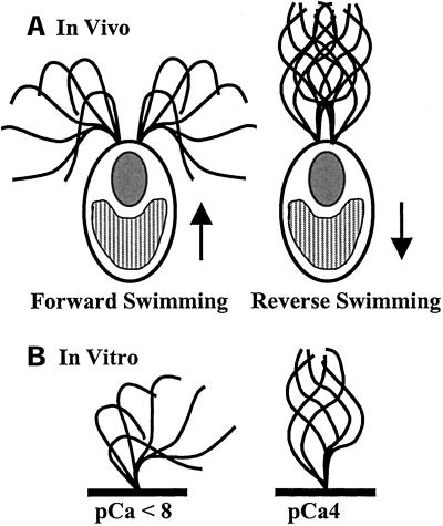
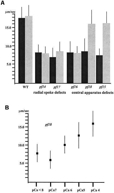
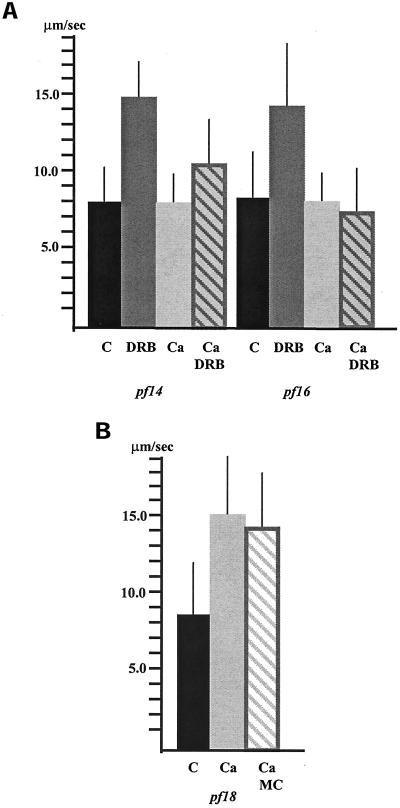
Ciliary and flagellar motility is regulated by changes in intraflagellar calcium. However, the molecular mechanism by which calcium controls motility is unknown. We tested the hypothesis that calcium regulates motility by controlling dynein-driven microtubule sliding and that the central pair and radial spokes are involved in this regulation. We isolated axonemes from Chlamydomonas mutants and measured microtubule sliding velocity in buffers containing 1 mM ATP and various concentrations of calcium. In buffers with pCa > 8, microtubule sliding velocity in axonemes lacking the central apparatus (pf18 and pf15) was reduced compared with that of wild-type axonemes. In contrast, at pCa4, dynein activity in pf18 and pf15 axonemes was restored to wild-type level. The calcium-induced increase in dynein activity in pf18 axonemes was inhibited by antagonists of calmodulin and calmodulin-dependent kinase II. Axonemes lacking the C1 central tubule (pf16) or lacking radial spoke components (pf14 and pf17) do not exhibit calcium-induced increase in dynein activity in pCa4 buffer. We conclude that calcium regulation of flagellar motility involves regulation of dynein-driven microtubule sliding, that calmodulin and calmodulin-dependent kinase II may mediate the calcium signal, and that the central apparatus and radial spokes are key components of the calcium signaling pathway.


Figures

Figure 1
Figure 1
(a) In vivo, Chlamydomonas cells normally swim forward, toward the light, with an asymmetric, ciliary waveform. During the photophobic response, bright light induces a shift from an asymmetric waveform to a symmetric, flagellar waveform, and the cells swim in reverse. The arrows indicate swimming direction (for example, see Ringo, 1967 and Ruffer and Nultsch, 1985). (b) This change in waveform can be induced in vitro. Isolated axonemes lacking membranes and soluble flagellar matrix components beat with an asymmetric waveform in buffers of pCa < 8 and beat with a symmetric waveform in buffers of pCa4. (Waveform traces adapted from Brokaw and Luck, 1985.)
Figure 2
Figure 2
(a) Microtubule sliding velocities in wild-type and mutant axonemes in low calcium (pCa > 8, black bars) and high calcium (pCa4, gray bars) buffers. Sliding velocities in wild-type, radial spoke–defective (pf14 and pf17), and C1 central tubule–defective (pf16) axonemes in low calcium buffer are not significantly different from those in high calcium buffer. In contrast, sliding velocities in axonemes completely lacking the central apparatus (pf15 and pf18) in high calcium buffer are significantly increased from those in low calcium buffer (p < 0.001; Student's t test). All bars represent the mean of >60 measurements ± SD from a minimum of three experiments. (b) Microtubule sliding velocity in pf18 axonemes in microtubule sliding buffer with varying concentrations of free calcium. As the concentration of free calcium increases, microtubule sliding velocity in pf18 axonemes increases to nearly wild-type level. Each point represents the mean of >40 measurements ± SD from two experiments.
Figure 3
Figure 3
(a) Microtubule sliding velocities in pf14 and pf16 axonemes. Dynein-driven microtubule sliding velocities in axonemes isolated from the radial spoke defective mutant pf14 and central apparatus defective mutant pf16 significantly increase (p < 0.001; Student's t test) upon the addition of DRB compared with control, untreated axonemes in low calcium buffer (C = pCa > 8). In high calcium buffer, (Ca = pCa4) DRB is less effective at increasing microtubule sliding velocity in pf14 axonemes and is completely ineffective at increasing sliding velocity in pf16 axonemes. (b) The increase in sliding velocity observed for pf18 axonemes in high calcium buffer is not inhibited by the addition of microcystin (MC). All bars represent the mean ± SD. N ≥ 60 from a minimum of three experiments.
Figure 4
Figure 4
(a) The addition of calmodulin inhibitors blocks the calcium-induced increase in sliding velocity observed for pf18 axonemes in pCa4 buffer (Ca). The sliding velocity in pf18 axonemes incubated in pCa4 buffer with either the calmodulin-binding domain peptide (CBD, 60.0 μM) or the calmodulin inhibitory peptide (CIP, 60.0 μM) is not significantly different from that in pf18 axonemes incubated in low calcium buffer. Sliding velocity in pf18 axonemes in pCa4 buffer incubated with the control peptide (CIPc, 60.0 μM) for the calmodulin inhibitory peptide is not significantly different from that in pf18 axonemes in pCa4 buffer alone. All bars represent the mean of >60 measurements ± SD from a minimum of three experiments. (b) Microtubule sliding velocity in pf18 axonemes in microtubule sliding buffer with varying concentrations of the calmodulin binding domain peptide (CBD) inhibitor. Increasing concentration of CBD decreases sliding velocity in pf18 axonemes in high calcium buffer. Each point represents the mean of >40 measurements ± SD from two experiments.
Figure 5
Figure 5
(a) The addition of CaM-KII inhibitors blocks the calcium-induced increase in sliding velocity observed for pf18 axonemes in pCa4 buffer (Ca). The sliding velocity in pf18 axonemes incubated in pCa4 buffer with either the compound KN-93 (1.0 μM) or the autocamtide-2 related inhibitory peptide (AIP, 3.0 μM) is not significantly different from that in pf18 axonemes incubated in low calcium buffer. Sliding velocity in pf18 axonemes in pCa4 buffer incubated with the control compound KN-92 (1.0 μM) is not significantly different from that in pf18 axonemes in pCa4 buffer alone. All bars represent the mean of >60 measurements ± SD from a minimum of three experiments. (b) Microtubule sliding velocity in pf18 axonemes in pCa4 microtubule sliding buffer with varying concentrations of the autocamtide-2 related inhibitory peptide (AIP). Increasing concentration of AIP decreases sliding velocity in pf18 axonemes in high calcium buffer. Each point represents the mean of >40 measurements ± SD from two experiments.

References

    1. Bannai H, Yoshimura M, Takahashi K, Shingyoji C. Calcium regulation of microtubule sliding in reactivated sea urchin sperm flagella. J Cell Sci. 2000;113:831–839. - PubMed
    1. Bessen M, Fay RB, Witman GB. Calcium control of waveform in isolated flagellar axonemes of. Chlamydomonas. J Cell Biol. 1980;86:446–455. - PMC - PubMed
    1. Bonini NM, Evans TC, Miglietta LA, Nelson DL. The regulation of ciliary motility in Paramecium by Ca2+ and cyclic nucleotides. Adv Second Messenger Phosphoprot Res. 1991;23:227–272. - PubMed
    1. Brokaw CJ. Calcium-induced asymmetrical beating of Triton-demembranated sea urchin sperm flagella. J Cell Biol. 1979;82:401–411. - PMC - PubMed
    1. Brokaw CJ. Calcium sensors in sea urchin sperm flagella. Cell Motil Cytoskelet. 1991;18:123–130. - PubMed

Publication types

LinkOut - more resources