

Convergent evolution sheds light on the anti-beta -elimination mechanism common to family 1 and 10 polysaccharide lyases
- PMID: 12221284
- PMCID: PMC129399
- DOI: 10.1073/pnas.182431199
Convergent evolution sheds light on the anti-beta -elimination mechanism common to family 1 and 10 polysaccharide lyases
Abstract
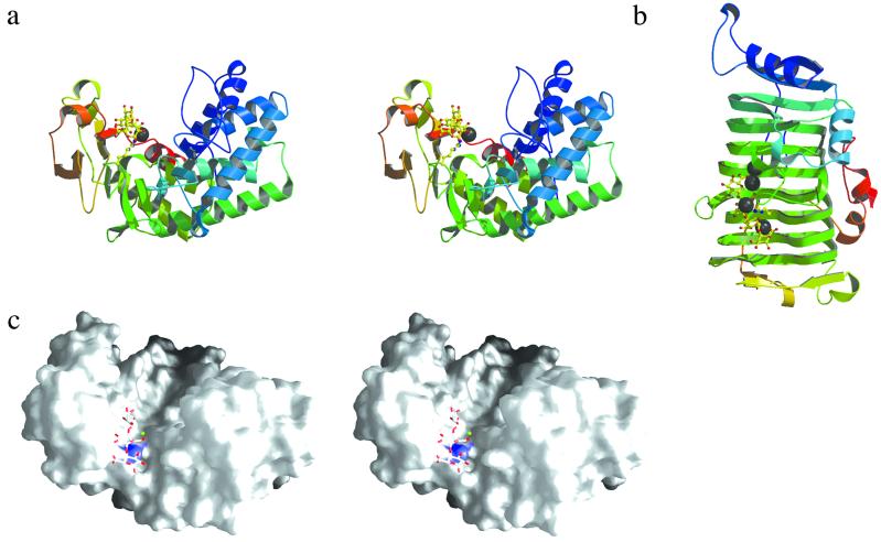
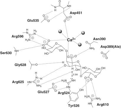
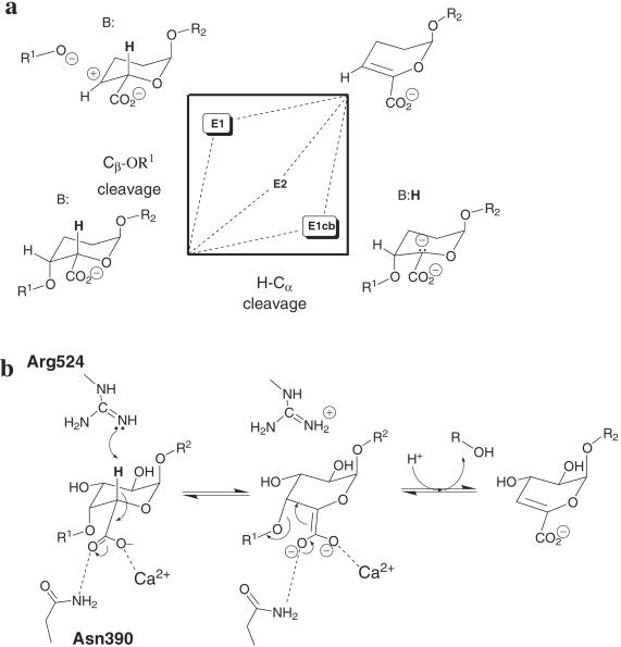
Enzyme-catalyzed beta-elimination of sugar uronic acids, exemplified by the degradation of plant cell wall pectins, plays an important role in a wide spectrum of biological processes ranging from the recycling of plant biomass through to pathogen virulence. The three-dimensional crystal structure of the catalytic module of a "family PL-10" polysaccharide lyase, Pel10Acm from Cellvibrio japonicus, solved at a resolution of 1.3 A, reveals a new polysaccharide lyase fold and is the first example of a polygalacturonic acid lyase that does not exhibit the "parallel beta-helix" topology. The "Michaelis" complex of an inactive mutant in association with the substrate trigalacturonate/Ca2+ reveals the catalytic machinery harnessed by this polygalacturonate lyase, which displays a stunning resemblance, presumably through convergent evolution, to the tetragalacturonic acid complex observed for a structurally unrelated polygalacturonate lyase from family PL-1. Common coordination of the -1 and +1 subsite saccharide carboxylate groups by a protein-liganded Ca2+ ion, the positioning of an arginine catalytic base in close proximity to the alpha-carbon hydrogen and numerous other conserved enzyme-substrate interactions, considered in light of mutagenesis data for both families, suggest a generic polysaccharide anti-beta-elimination mechanism.
Figures

References
-
- Anderson V. E. (1998) in Comprehensive Biological Catalysis: A Mechanistic Reference, ed. Sinnott, M. (Academic, London), Vol. 2, pp. 115–133.
-
- Coutinho P. M. & Henrissat, B. (1999) in Recent Advances in Carbohydrate Engineering, eds. Gilbert, H. J., Davies, G. J., Svensson, B. & Henrissat, B. (R. Soc. Chem., Cambridge, U.K.), pp. 3–12.
Publication types
MeSH terms
Substances
Grants and funding
LinkOut - more resources
Full Text Sources
Miscellaneous
