Comparative Study
. 2002 Sep 17;99(19):12321-6.
doi: 10.1073/pnas.192449599. Epub 2002 Sep 9.

Inhibition of Fe-S cluster biosynthesis decreases mitochondrial iron export: evidence that Yfh1p affects Fe-S cluster synthesis

Affiliations
Comparative Study

Inhibition of Fe-S cluster biosynthesis decreases mitochondrial iron export: evidence that Yfh1p affects Fe-S cluster synthesis

Opal S Chen et al. Proc Natl Acad Sci U S A. .

Abstract

Decreased expression of Yfh1p in the budding yeast, Saccharomyces cerevisiae, and the orthologous human gene frataxin results in respiratory deficiency and mitochondrial iron accumulation. The absence of Yfh1p decreases mitochondrial iron export. We demonstrate that decreased expression of Nfs1p, the yeast cysteine desulfurase that plays a central role in Fe-S cluster synthesis, also results in mitochondrial iron accumulation due to decreased export of mitochondrial iron. In the absence of Yfh1p, activity of Fe-S-containing enzymes (aconitase, succinate dehydrogenase) is decreased, whereas the activity of a non-Fe-S-containing enzyme (malate dehydrogenase) is unaffected. Aconitase protein was abundant even though the activity of aconitase was decreased in both aerobic and anaerobic conditions. These results demonstrate a direct role of Yfh1p in the formation of Fe-S clusters and indicate that mitochondrial iron export requires Fe-S cluster biosynthesis.


Figures

Fig 1.
Fig 1.
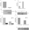
Expression of Yfh1p affects FET3 transcription. Wild-type cells (transformed with a control vector) were grown in CM media and then transferred to CM media containing the iron-chelator BPS at concentrations high enough to chelate all available iron. Overnight cultures of METYFH1 cells grown in CM with methionine (330 μg/ml) were transferred to BPS-containing media in the presence (“off to off”) or absence (“off to on”) of methionine. At selected times cells were harvested, mRNA was isolated and analyzed by S1 nuclease analysis for both FET3 and CMD1 transcripts.
Fig 2.
Fig 2.
Expression of Nfs1p affects mitochondrial iron export and FET3 transcription. (A) METNSF1 and METYFH1 cells were grown overnight in the presence of methionine. The cells were incubated for 10 min with 59Fe in the presence of ascorbate and then incubated in 59Fe-free media for 12 h in the presence or absence of methionine. The cells were sphereoplasted, homogenized, and the membrane fraction applied to an Iodoxianol gradient. Radioactivity was determined in each fraction and was plotted as a percentage of total cell radioactivity. The mitochondria are found in fractions 3–9. (B) The experimental design was as described in the legend to Fig. 1 except that METNFS1 was studied.
Fig 3.
Fig 3.
Yfh1p depletion affects the activity of aconitase. A Triton X-100 mitochondrial extract from wild-type, rho0, and Δyfh1 strains was assayed for aconitase activity (A), succinate dehydrogenase activity (B), and malate dehydrogenase activity (C). Aconitase and porin protein levels were determined by Western blot analysis on isolated mitochondria (D). The degradation of aconitase protein on Western blot analysis is denoted by an asterisk.
Fig 4.
Fig 4.
Aconitase activity in METYFH1 cells overexpressing ACO1 and CCC1. Aconitase activity (Upper) and protein (Lower) were assayed in METYFH1 cells (A), in METYFH1 cells transformed with a high-copy ACO1 plasmid or vector (B), in METYFH1 cells transformed with a high-copy CCC1 plasmid (C), or with both high-copy ACO1 and CCC1 plasmids (D). The cells were grown in the presence (Yfh1p “off”) or absence (Yfh1p “on”) of methionine for 15 h, a total cell extract prepared, and aconitase activity or protein determined. The degradation of aconitase protein on Western blot analysis is denoted by an asterisk.
Fig 5.
Fig 5.
Yfh1p depletion affects aconitase activity in cells grown under anaerobic conditions. Control cells (wild-type and wild-type rho0 cells) and Δyfh1 cells were grown in yeast extract peptone dextrose media under anaerobic conditions for 3 days. Mitochondria were isolated and a Triton X-100-soluble fraction was used to determine the activities of aconitase (A), malate dehydrogenase (B), and succinate dehydrogenase (C). (The increase in specific activities in this figure, compared with Fig. 4, is due to use of the more purified Triton X-100 extract to assay enzyme activities.) (D) The abundance of aconitase in isolated mitochondria was determined by Western blot analysis. The level of the mitochondrial protein porin was determined by Western blot analysis to demonstrate equal loading of total mitochondrial protein.
Fig 6.
Fig 6.
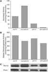
Yfh1p depletion affects aconitase in cells overexpressing ACO1 grown under anaerobic conditions. Both wild-type rho0 cells and Δyfh1 cells transformed with a high-copy ACO1 plasmid (pACO1) or a control vector (V) were grown in CM media under anaerobic conditions for 3 days. Mitochondria were isolated, and the activities of aconitase (A) and malate dehydrogenase (B) were determined in the Triton X-100-soluble mitochondrial fraction. (C) Aconitase and porin protein levels were determined by Western blot analysis.

References

    1. Campuzano V., Montermini, L., Molto, M. D., Pianese, L., Cossee, M., Cavalcanti, F., Monros, E., Rodius, F., Duclos, F., Monticelli, A., et al. (1996) Science 271, 1423-1427. - PubMed
    1. Babcock M., de Silva, D., Oaks, R., Davis-Kaplan, S., Jiralerspong, S., Montermini, L., Pandolfo, M. & Kaplan, J. (1997) Science 276, 1709-1172. - PubMed
    1. Radisky D. C., Babcock, M. C. & Kaplan, J. (1999) J. Biol. Chem. 274, 4497-4499. - PubMed
    1. Foury F. (1999) FEBS Lett. 456, 281-284. - PubMed
    1. Chen O. S. & Kaplan, J. (2000) J. Biol. Chem. 275, 7626-7632. - PubMed

Publication types

MeSH terms