

Age-related total gray matter and white matter changes in normal adult brain. Part I: volumetric MR imaging analysis
- PMID: 12223373
- PMCID: PMC7976241
Age-related total gray matter and white matter changes in normal adult brain. Part I: volumetric MR imaging analysis
Abstract
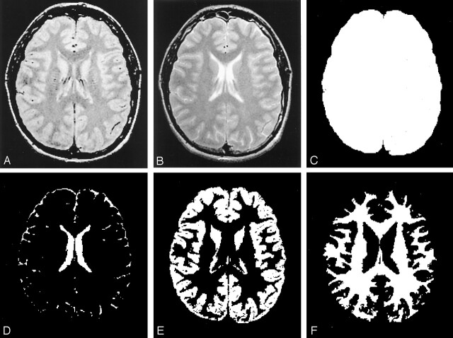
Background and purpose: A technique of segmenting total gray matter (GM) and total white matter (WM) in human brain is now available. We investigated the effects of age and sex on total fractional GM (%GM) and total fractional WM (%WM) volumes by using volumetric MR imaging in healthy adults.
Methods: Fifty-four healthy volunteers (22 men, 32 women) aged 20-86 years underwent dual-echo fast spin-echo MR imaging. Total GM, total WM, and intracranial space volumes were segmented by using MR image-based computerized semiautomated software. Volumes were normalized as a percentage of intracranial volume (%GM and %WM) to adjust for variations in head size. Age and sex effects were then assessed.
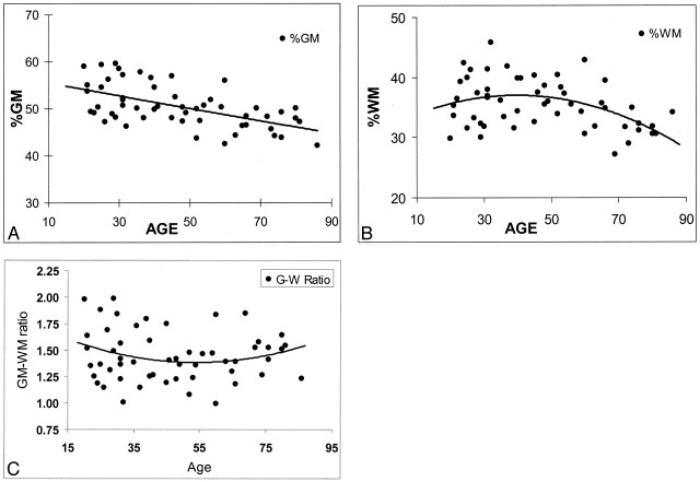
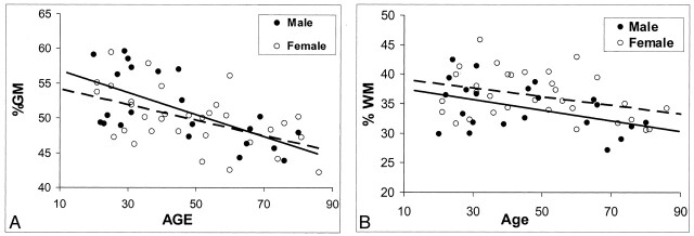
Results: Both %GM and %WM in the intracranial space were significantly less in older subjects (> or =50 years) than in younger subjects (<50 years) (P <.0001 and P =.02, respectively). Consistently, %GM decreased linearly with age, beginning in the youngest subjects. %WM decreased in a quadratic fashion, with a greater rate beginning only in adult midlife. Although larger GM volumes were observed in men before adjustments for cranium size, no significant differences in %GM or %WM were observed between the sexes.
Conclusion: GM volume loss appears to be a constant, linear function of age throughout adult life, whereas WM volume loss seems to be delayed until middle adult life. Both appear to be independent of sex. Quantitative analysis of %GM and %WM volumes can improve our understanding of brain atrophy due to normal aging; this knowledge may be valuable in distinguishing atrophy of disease patterns from characteristics of the normal aging process.
Figures
Comment in
-
Standardized calculation of brain parenchymal fraction: an approach to objective assessment of cerebral atrophy.AJNR Am J Neuroradiol. 2003 Aug;24(7):1492-3; author reply 1493. AJNR Am J Neuroradiol. 2003. PMID: 12917155 Free PMC article. No abstract available.
References
-
- Biegon A, Eberling JL, Richardson BC, et al. Human corpus callosum in aging and Alzheimer’s disease: a magnetic resonance imaging study. Neurobiol Aging 1994;15:393–397 - PubMed
-
- Giedd JN, Vaituzis AC, Hamburger SD, et al., Quantitative MRI of the temporal lobe, amygdala, and hippocampus in normal human development: ages 4–18 years. J Comp Neuro 1996;366:223–230 - PubMed
-
- Daigneault S, Braun CMJ, Whitaker HA. Early effects of normal aging on preservative and nonpreservative prefrontal measures. Dev Neuropsychol 1992;8:99–114
-
- Convit A, de Leon MJ, Hoptman MJ, Tarshish C, De Santi S, Rusinek H. Age-related changes in brain: I. Magnetic resonance imaging measures of temporal lobe volumes in normal subjects. Psychiatr Q 1995;66:343–355 - PubMed
-
- Luft AR, Skalej M, Schulz JB, et al. Patterns of age-related shrinkage in cerebellum and brainstem observed in vivo using three-dimensional MRI volumetry. Cerebr Cortex 1999;9:712–721 - PubMed
Publication types
MeSH terms
Grants and funding
LinkOut - more resources
Full Text Sources
Other Literature Sources
Medical