

Proton MR spectroscopy of tumefactive demyelinating lesions
- PMID: 12223381
- PMCID: PMC7976261
Proton MR spectroscopy of tumefactive demyelinating lesions
Abstract
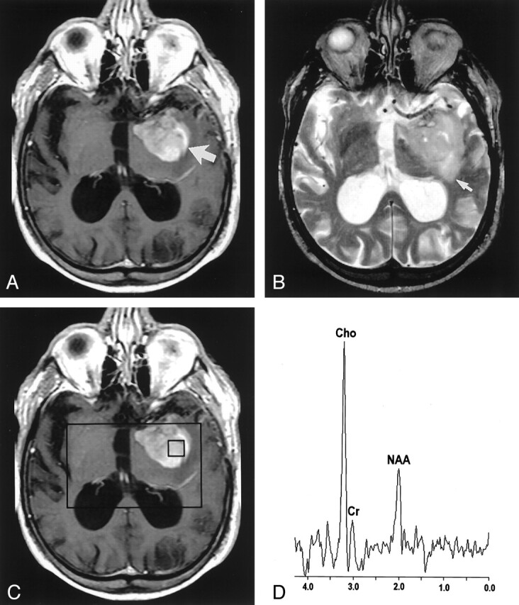
Background and purpose: Tumefactive demyelinating lesions (TDLs) can simulate intracranial neoplasms in clinical presentation and MR imaging appearance, and surgical biopsy is often performed in suspected tumors. Proton MR spectroscopy has been applied in assessing various intracranial diseases and is increasingly used in diagnosis and clinical management. Our purpose was to determine if multivoxel proton MR spectroscopy can be used to differentiate TDLs and high-grade gliomas.
Methods: Conventional MR images, proton MR spectra, and medical records were retrospectively reviewed in six patients with TDLs diagnosed by means of biopsy or by documented clinical improvement, with or without supporting laboratory testing and follow-up imaging. Proton MR spectra of 10 high-grade gliomas with similar conventional MR imaging appearances were used for comparison. In contrast-enhancing, central, and perilesional areas of each lesion, peak heights of N-acetylaspartate (NAA), choline (Cho), and creatine (Cr) were measured and the lactate peak noted. Cho/Cr and NAA/Cr ratios of corresponding regions in TDLs and gliomas were compared.
Results: No significant differences in mean Cho/Cr ratios were found in the corresponding contrast-enhancing, central, or perilesional areas of TDLs and gliomas. The mean central-region NAA/Cr ratio in gliomas was significantly lower than that of TDLs, but mean NAA/Cr ratios in other regions were not significantly different. A lactate peak was identified in four of six TDLs and three of 10 gliomas.
Conclusion: In the cases examined, the NAA/Cr ratio in the central region of TDLs and high-grade gliomas differed significantly. However, overall metabolite profiles of both lesions were similar; this finding emphasizes the need for the cautious interpretation of spectroscopic findings.
Figures

References
-
- Mastrostefano R, Occhipinti E, Bigotti G, et al. Multiple sclerosis plaque simulating cerebral tumor: case report and review of the literature. Neurosurgery 1987;21:244–246 - PubMed
-
- Hunter SB, Ballinger WE Jr, Rubin JJ. Multiple sclerosis mimicking primary brain tumor. Arch Pathol Lab Med 1987;111:464–468 - PubMed
-
- Giang DW, Poduri KR, Eskin TA, et al. Multiple sclerosis masquerading as a mass lesion. Neuroradiology 1992;34:150–154 - PubMed
-
- Silva HC, Callegaro D, Marchiori PE, et al. Magnetic resonance imaging in five patients with a tumefactive demyelinating lesion in the central nervous system. Arq Neuropsiquiatr 1999;57:921–926 - PubMed
-
- Kalyan-Raman UP, Garwacki DJ, Elwood PW. Demyelinating disease of corpus callosum presenting as glioma on magnetic resonance scan: a case documented with pathological findings. Neurosurgery 1987;21:247–250 - PubMed
Publication types
MeSH terms
Substances
LinkOut - more resources
Full Text Sources
Medical