

Case Reports
MR changes after acute cyanide intoxication
Affiliations
- PMID: 12223384
- PMCID: PMC7976263
Item in Clipboard
Case Reports
MR changes after acute cyanide intoxication
AJNR Am J Neuroradiol.
2002 Sep.
Abstract
We describe MR changes that occurred 3 and 6 weeks after a suicide attempt with cyanide. The toxicity of cyanide causes damage, primarily to the basal ganglia, and those changes were visible as altered signal intensity on the first MR images. Extensive areas of hemorrhagic necrosis were seen 6 weeks later. Our case shows pseudolaminar necrosis along the central cerebral cortex 3 weeks after cyanide poisoning, showing that the sensorimotor cortex is also a site for toxic necrosis because of its high oxygen dependency.
Figures
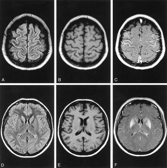
Follow-up MR images obtained 3 weeks after cyanide poisoning. A, Fluid-attenuated inversion recovery (9000/110 [TR/TE]) image. Discrete hyperintense linear signals can be seen along the sensorimotor cortex. B, T1-weighted (550/14) image obtained before the administration of contrast agent. C, T1-weighted (715/17) image obtained after the administration of contrast agent. Massive contrast enhancement can be seen along the central region, consistent with pseudolaminar necrosis. D, Fluid-attenuated inversion recovery (9000/110) image. Discrete hyperintense signals can be seen in the caudate nuclei bilaterally. E, Unenhanced T1-weighted (550/14) image shows hyperintense signals in the caudate nuclei bilaterally, consistent with hemorrhage. F, T1-weighted (715/17) image obtained after administration of contrast agent. Massive contrast enhancement can be seen in the lentiform nuclei and the caudate nuclei bilaterally.
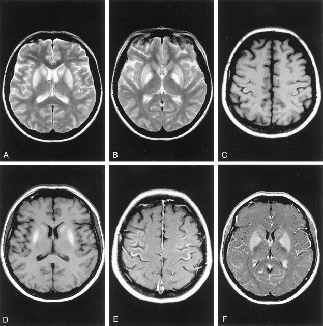
Follow-up MR images obtained 6 weeks after cyanide poisoning. A and B, T2-weighted (2660/90) images. The hyperintense signal changes in the basal ganglia are considerably more pronounced. C and D, T1-weighted (550/14) unenhanced images. Hemorrhagic necrosis can be seen in the sensorimotor cortex (C) and the basal ganglia (D). E and F, T1-weighted (715/17) images obtained after administration of contrast agent. Pronounced contrast enhancement can be seen along the central cortex (E) and the basal ganglia (F).
References
-
- Heiss WD, Würker M. Value of functional imaging in Parkinson’s disease and related movement disorders [in German]. Nervenarzt 1999;70[suppl 1]:S2–S10 - PubMed
-
- Moeschlin S. Klinik und Therapie der Vergiftungen. 6th ed. Stuttgart: Georg Thieme Verlag;1980. :252–256
-
- Quadbeck G, Ule G. Die toxischen Stoffgruppen. II. Zyanwasserstoff und Zyanide. In: Berlet H, Noetzel N, Quadbeck G, Scholz W, Schmitt HP, Ule G, Ule VG, eds. Pathologie des Nervensystems II.Berlin: Springer-Verlag;1983. :291–299
-
- Henschler D. Wichtige Gifte und Vergiftungen. In: Forth W, Henschler D, Rummel W, eds. Allgemeine und spezielle Pharmakologie und Toxikologie.5th ed. Wissenschaftsverlag: Mannheim1987. :751–752
-
- Borgohain R, Singh AK, Radhakrishna H, Rao VC, Mohandas S. Delayed onset generalised dystonia after cyanide poisoning. Clin Neurol Neurosurg 1995;97:213–215 - PubMed
Publication types
MeSH terms
Substances
LinkOut - more resources
Full Text Sources
Medical