. 2002 Sep 15;22(18):7873-8.
doi: 10.1523/JNEUROSCI.22-18-07873.2002.

Non-Fc-mediated mechanisms are involved in clearance of amyloid-beta in vivo by immunotherapy

Affiliations

Non-Fc-mediated mechanisms are involved in clearance of amyloid-beta in vivo by immunotherapy

Brian J Bacskai et al. J Neurosci. .

Abstract

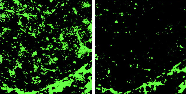
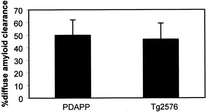
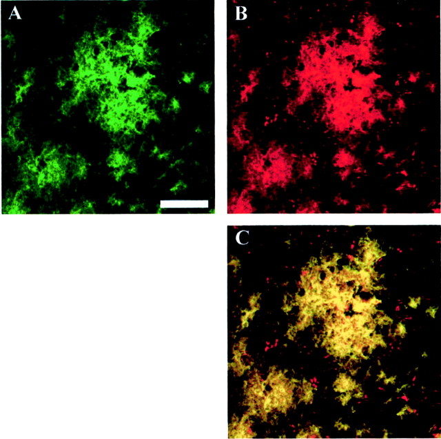
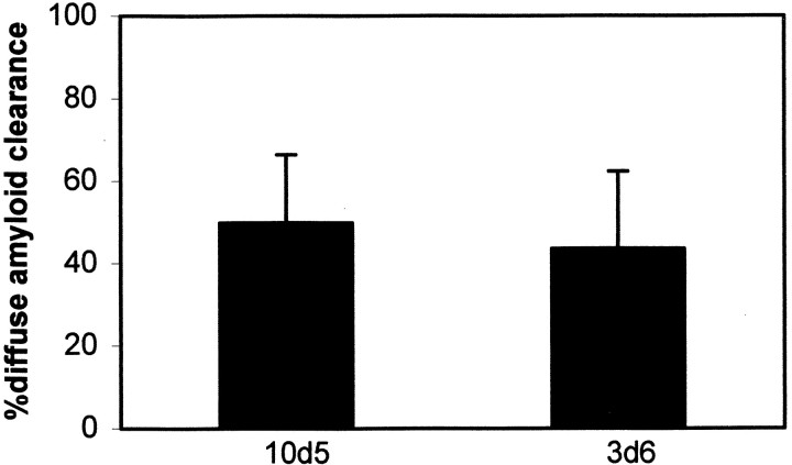
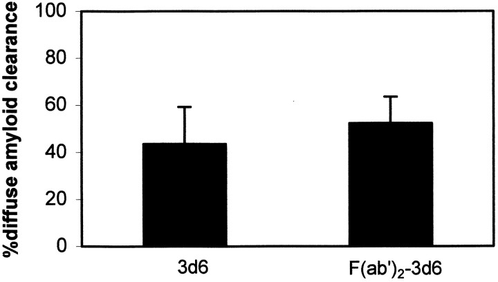
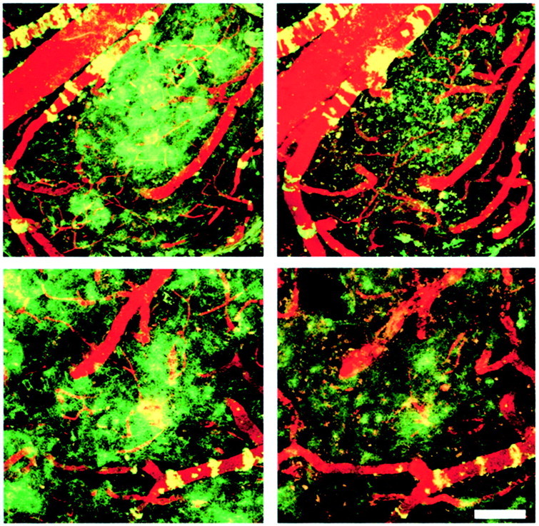
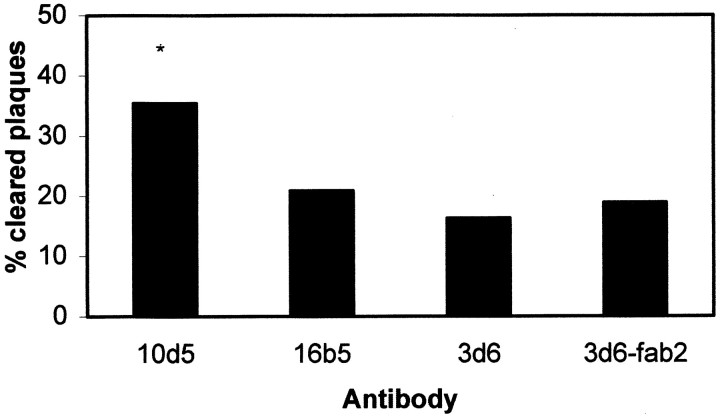
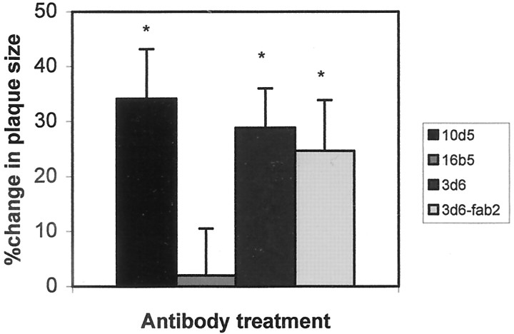
Transgenic (Tg) mouse models overexpressing amyloid precursor protein (APP) develop senile plaques similar to those found in Alzheimer's disease in an age-dependent manner. Recent reports demonstrated that immunotherapy is effective at preventing or removing amyloid-beta deposits in the mouse models. To characterize the mechanisms involved in clearance, we used antibodies of either IgG1 (10d5) or IgG2b (3d6) applied directly to the brains of 18-month-old Tg2576 or 20-month-old PDAPP mice. Both 10d5 and 3d6 led to clearance of 50% of diffuse amyloid deposits in both animal models within 3 d. Fc receptor-mediated clearance has been shown to be important in an ex vivo assay showing antibody-mediated clearance of plaques by microglia. We now show, using in vivo multiphoton microscopy, that FITC-labeled F(ab')2 fragments of 3d6 (which lack the Fc region of the antibody) also led to clearance of 45% of the deposits within 3 d, similar to the results obtained with full-length 3d6 antibody. This result suggests that direct disruption of plaques, in addition to Fc-dependent phagocytosis, is involved in the antibody-mediated clearance of amyloid-beta deposits in vivo. Dense-core deposits that were not cleared were reduced in size by approximately 30% with full-length antibodies and F(ab')2 fragments 3 d after a topical treatment. Together, these results indicate that clearance of amyloid deposits in vivo may involve, in addition to Fc-dependent clearance, a non-Fc-mediated disruption of plaque structure.


Figures

Fig. 1.
Fig. 1.
Topical antibody application leads to clearance of diffuse amyloid-β in Tg2576 mice. These images are projections of three-dimensional volumes from the cortex of a representative Tg2576 mouse treated with FITC-labeled 10d5 antibody. The images were acquired in the anesthetized mouse using multiphoton microscopy. Theleft shows labeled amyloid deposits at the initial application of antibody. Numerous diffuse deposits as well as amyloid angiopathy can be seen. The right is the same volume 3 d later, labeled with 3d6 antibody, which recognizes amyloid-β independently of 10d5. A majority of the amyloid-β deposits have been cleared in this 3 d period.
Fig. 2.
Fig. 2.
10d5 antibody is equally effective at clearing diffuse amyloid-β deposits in both PDAPP and Tg2576 transgenic mouse models. These plots represent the percentage of amyloid clearance in 3 d from paired volumes of cortex within the same animals after 3 d. A single application of antibody to the cortex was given at day 0. Approximately one-half of the diffuse deposits are cleared under these conditions. These results are the means ± SE forn = 9–15 sites from at least three animals in each group. Percentage of clearance ranged from 91.2 to −7.9% in PDAPP mice and 82.9 to 4.5% in Tg2576 mice. Not statistically different; Student's t test.
Fig. 3.
Fig. 3.
10d5 and 3d6 antibodies colocalize at noncompetitive binding sites on amyloid-β plaques. Paraformaldehyde-fixed tissue sections from an 18-month-old Tg2576 mouse brain were treated sequentially with FITC-labeled 10d5 antibodies, followed by rhodamine-labeled 3d6 antibodies. Images were obtained with a Bio-Rad confocal microscope using 488 nm excitation for FITC (A) and 568 nm excitation for rhodamine (B). A color-merged image (C) shows that 10d5 (green) and 3d6 (red) are both able to bind to the plaques and colocalize everywhere, producing a yellow color. Scale bar, 50 μm.
Fig. 4.
Fig. 4.
10d5 and 3d6 antibodies are equally effective at clearing diffuse amyloid-β deposits in the PDAPP mouse model after 3 d. 10d5 and 3d6 recognize the N-terminus residues 3–6 and 1–5, respectively, but their epitopes do not physically overlap. 10d5 (IgG1) and 3d6 (IgG2b) are different isotypes as well, suggesting that clearance in vivo is not specific to any one class of antibody. These data represent n = 9 sites from four animals. Not statistically different; Student's ttest.
Fig. 5.
Fig. 5.
F(ab′)2 fragments of 3d6 antibody are equally effective at leading to clearance of diffuse amyloid-β deposits after 3 d in the PDAPP mouse model. F(ab′)2fragments were purified and analyzed for Fc contamination by Western blot analysis and immunohistochemistry. No evidence for trace Fc was detected with either assay. Approximately one-half of the diffuse amyloid-β deposits were cleared 3 d after a single topical application to the cortex of the transgenic mice. Error bars represent means ± SE from n = 15 or 11 sites from four animals from each group. Not statistically different; Student's t test.
Fig. 6.
Fig. 6.
F(ab′)2 fragments of 3d6 antibody are equally effective at leading to clearance of diffuse amyloid-β deposits after 3 d in the PDAPP mouse model. These images are projections of three-dimensional volumes from the cortex of a representative PDAPP mouse treated with FITC-labeled 3d6 or 3d6-F(ab′)2 antibodies. The images were acquired in the anesthetized mouse using multiphoton microscopy. The left images shows labeled amyloid deposits at the initial application of antibody. Numerous diffuse deposits as well as amyloid angiopathy can be seen, pseudocolored green. A fluorescent angiogram (red) fills the blood vessels in each imaging session to facilitate image alignment. The right images are the same volumes 3 d later, immediately after application of labeled 10d5 antibody, which recognizes amyloid-β independently of 3d6. A majority of the amyloid-β deposits have been cleared in this 3 d period with both full-length (top row) and F(ab′)2 fragments (bottom row) of antibody. Scale bar, 100 μm.
Fig. 7.
Fig. 7.
Dense-core plaques are cleared with 10d5 after 3 d in PDAPP mice. thioS-positive plaques were identified and counted in animals at the initial imaging–treatment session. In the subsequent session, after 3 d, the presence or absence of the individual plaques was scored in the identical locations within each animal, using the fluorescent angiogram as both a three-dimensional fiduciary marker, as well as a positive control for imaging quality. Two independent observers scored the presence or absence of identifiable plaques in the PDAPP mice. 16b5 is a fluorescently labeled control antibody that recognizes human tau. 3d6 and F(ab′)2fragments of 3d6 did not lead to appreciable clearance of thioS plaques after 3 d, whereas 10d5 led to the removal of 35% of identified plaques. Bars represent the percentage of identified plaques that were not found after 3 d. At least 45 plaques from 8–12 sites in four animals were scored for each group. *p < 0.05 indicates statistically significant byt test.
Fig. 8.
Fig. 8.
Remaining dense-core plaques in PDAPP mice are reduced in size 3 d after treatment with anti-amyloid-β antibodies. The identified plaques that were imaged in the first imaging session and remained after 3 d after a single antibody treatment were measured as described previously (Christie et al., 2001). Plaques that were cleared completely would have been measured as 100% change in size but are not included in this analysis. 10d5, 3d6, and F(ab′)2 fragments of 3d6 were equally effective at reducing the size of remaining dense-core plaques compared with control antibody 16b5. Error bars are mean ± SE from at least 12 plaques in four animals from each group. *p < 0.05 indicates statistically significant by Student's ttest.

References

    1. Bacskai BJ, Kajdasz ST, Christie RH, Carter C, Games D, Seubert P, Schenk D, Hyman BT. Imaging of amyloid-beta deposits in brains of living mice permits direct observation of clearance of plaques with immunotherapy. Nat Med. 2001;7:369–372. - PubMed
    1. Bard F, Cannon C, Barbour R, Burke RL, Games D, Grajeda H, Guido T, Hu K, Huang J, Johnson-Wood K, Khan K, Kholodenko D, Lee M, Lieberburg I, Motter R, Nguyen M, Soriano F, Vasquez N, Weiss K, Welch B, Seubert P, Schenk D, Yednock T. Peripherally administered antibodies against amyloid beta-peptide enter the central nervous system and reduce pathology in a mouse model of Alzheimer disease. Nat Med. 2000;6:916–919. - PubMed
    1. Bornemann KD, Wiederhold KH, Pauli C, Ermini F, Stalder M, Schnell L, Sommer B, Jucker M, Staufenbiel M. Abeta-induced inflammatory processes in microglia cells of APP23 transgenic mice. Am J Pathol. 2001;158:63–73. - PMC - PubMed
    1. Christie RH, Bacskai BJ, Zipfel WR, Williams RM, Kajdasz ST, Webb WW, Hyman BT. Growth arrest of individual senile plaques in a model of Alzheimer's disease observed by in vivo multiphoton microscopy. J Neurosci. 2001;21:858–864. - PMC - PubMed
    1. DeMattos RB, Bales KR, Cummins DJ, Dodart JC, Paul SM, Holtzman DM. Peripheral anti-A beta antibody alters CNS and plasma A beta clearance and decreases brain A beta burden in a mouse model of Alzheimer's disease. Proc Natl Acad Sci USA. 2001;98:8850–8855. - PMC - PubMed

Publication types

MeSH terms