. 2002 Sep 15;22(18):7991-8001.
doi: 10.1523/JNEUROSCI.22-18-07991.2002.

Calmodulin is an auxiliary subunit of KCNQ2/3 potassium channels

Affiliations

Calmodulin is an auxiliary subunit of KCNQ2/3 potassium channels

Hua Wen et al. J Neurosci. .

Abstract

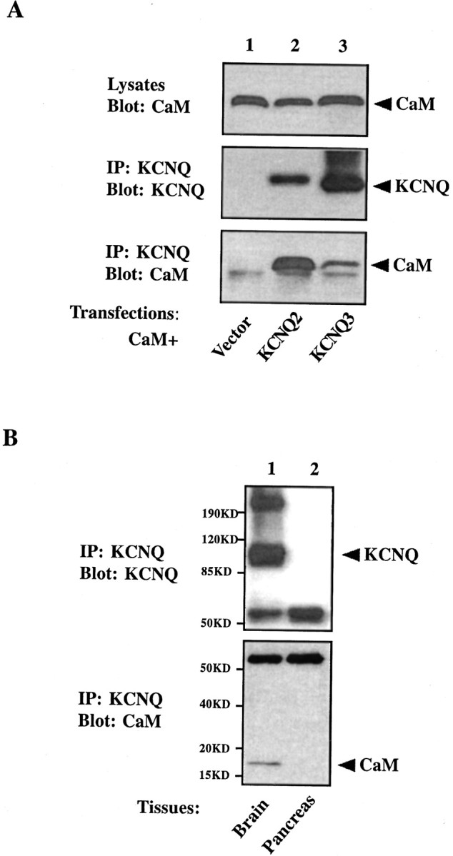
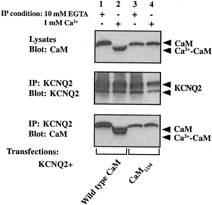
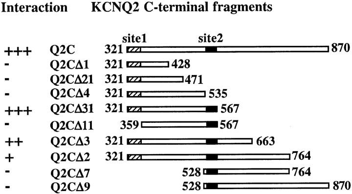
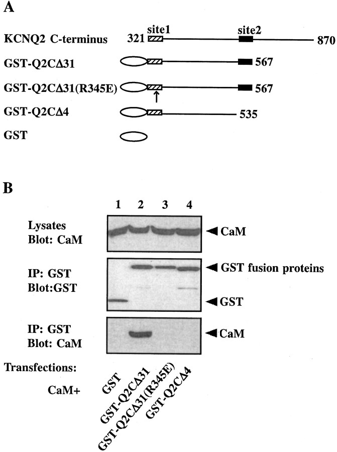
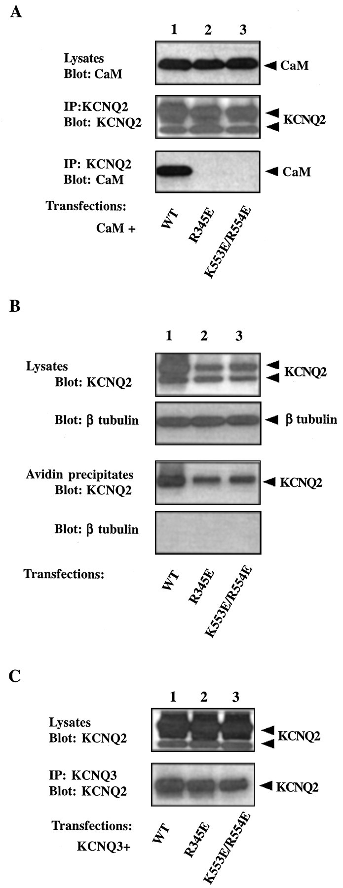
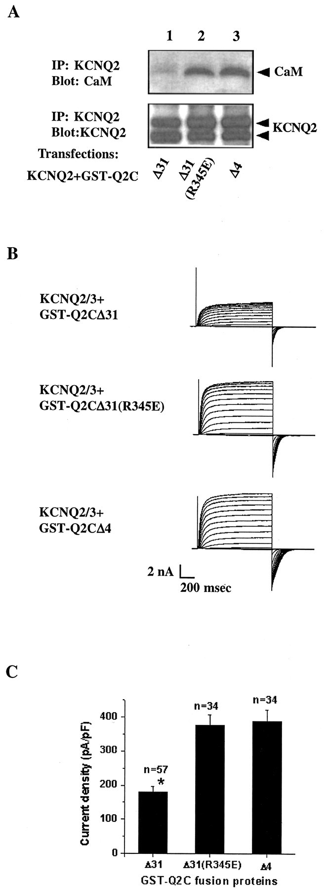
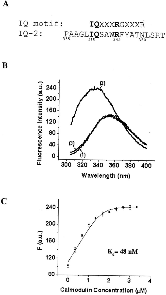
Calmodulin (CaM) was identified as a KCNQ2 and KCNQ3 potassium channel-binding protein, using a yeast two-hybrid screen. CaM is tethered constitutively to the channel, in the absence or presence of Ca2+, in transfected cells and also coimmunoprecipitates with KCNQ2/3 from mouse brain. The structural elements critical for CaM binding to KCNQ2 lie in two conserved motifs in the proximal half of the channel C-terminal domain. Truncations and point mutations in these two motifs disrupt the interaction. The first CaM-binding motif has a sequence that conforms partially to the consensus IQ motif, but both wild-type CaM and a Ca2+-insensitive CaM mutant bind to KCNQ2. The voltage-dependent activation of the KCNQ2/3 channel also shows no Ca2+ sensitivity, nor is it affected by overexpression of the Ca2+-insensitive CaM mutant. On the other hand, KCNQ2 mutants deficient in CaM binding are unable to generate detectable currents when coexpressed with KCNQ3 in CHO cells, although they are expressed and targeted to the cell membrane and retain the ability to assemble with KCNQ3. A fusion protein containing both of the KCNQ2 CaM-binding motifs competes with the full-length KCNQ2 channel for CaM binding and decreases KCNQ2/3 current density in CHO cells. The correlation of CaM binding with channel function suggests that CaM is an auxiliary subunit of the KCNQ2/3 channel.


Figures

Fig. 1.
Fig. 1.
CaM coimmunoprecipitates with KCNQ2 and KCNQ3 channels. A, tsA 201 cells were transfected with HA-tagged CaM together with one of following constructs: vector (lane 1), V5-tagged KCNQ2 (lane 2), or V5-tagged KCNQ3 (lane 3). Cell lysates were probed for the expression of CaM with the anti-HA antibody (top panel). Channel immunoprecipitates were probed with the anti-V5 antibody to confirm the precipitation of channels (middle panel) and were probed with the anti-HA antibody to detect CaM pulled down with the channels (bottom panel). CaM is present in a KCNQ channel immunoprecipitate, but not in that from vector-transfected cells.B, CaM copurifies with KCNQ2 and KCNQ3 channels from mouse brain. KCNQ2 and KCNQ3 channels were immunopurified from the crude membrane fraction of mouse brain (lane 1), but not pancreas (lane 2), with anti-KCNQ2/3 antibody-coupled Sepharose beads (top panel). CaM is detected in the channel immunoprecipitates from brain, but not pancreas, when probed with a specific monoclonal antibody against CaM (bottom panel).
Fig. 2.
Fig. 2.
Both Ca2+-CaM and apo-CaM coimmunoprecipitate with KCNQ2. tsA 201 cells were transfected with V5-tagged KCNQ2 together with one of two HA-tagged CaM constructs: wild type (lanes 1, 2) or Ca2+-insensitive mutant (CaM1234; lanes 3, 4). Cell lysates were probed for the expression of CaMs with the anti-HA antibody (top panel). KCNQ2 was precipitated with a polyclonal antibody against KCNQ2/3 in the presence of either 10 mm EGTA (lanes 1, 3) or 1 mm Ca2+ (lanes 2, 4), as indicated on the top of the blots. Channel immunoprecipitates were eluted with sample loading buffer containing 2 mm EGTA (for precipitates prepared in the presence of 10 mm EGTA) or 5 mmCa2+ (for precipitates prepared in the presence of 1 mm Ca2+) and were used for Western blot. Blots were probed for the channel by using anti-V5 antibody (middle panel) and for CaMs by using anti-HA antibody (bottom panel). A doublet usually shows up in the channel blot (middle panel) with longer exposure. Both bands represent overexpressed KCNQ2 protein because they are absent in vector-transfected cells. CaM is detected in all immunoprecipitates. Ca2+-loaded CaM (lane 2) shows greater mobility than the apo-CaM (CaM1234and wild-type CaM in the presence of EGTA) on the blots.
Fig. 3.
Fig. 3.
Truncation analysis reveals two sites in the KCNQ2 C-terminal domain that are necessary for CaM–KCNQ2 interaction. KCNQ2 C-terminal fragments (shown as boxes with amino acidnumbers) were tested against CaM in the two-hybrid system for LacZ reporter gene expression (positive interactions are indicated by plus signs). Further truncation from either end of Q2CΔ31 (amino acids 321–567) abolished the interaction. The locations of the two putative CaM-binding sites, site 1 (amino acids 321–358; hatched boxes) and site 2 (amino acids 536–567; filled boxes), are indicated in each fragment.
Fig. 4.
Fig. 4.
A GST fusion protein containing both binding sites coimmunoprecipitates with CaM. A, Schematic view of the fusion protein constructs. GST is shown as an oval. KCNQ2 C-terminal fragments are presented as lines with amino acid numbers. The locations of site 1 (hatched boxes) and site 2 (filled boxes) are indicated. The position of a mutation introduced in site 1, R345E, is indicated by an arrow.B, tsA 201 cells were transfected with HA-tagged CaM together with GST (lane 1), GST-Q2CΔ31 (lane 2), GST-Q2CΔ31(R345E) (lane 3), and GST-Q2CΔ4 (lane 4). Cell lysates were probed for the expression of CaM by using an anti-HA antibody (top panel). GST immunoprecipitates were probed for GST with anti-GST (middle panel) and for CaM with anti-HA (bottom panel). The fusion protein containing both sites, GST-Q2CΔ31, coimmunoprecipitates with CaM, whereas the one lacking site 2, GST-Q2CΔ4, does not. A single amino acid mutation (R345E) in site 1 also disrupts the interaction.
Fig. 5.
Fig. 5.
Properties of two KCNQ2 channels with mutations in CaM-binding sites. A, R345E and K553E/R554E KCNQ2 do not coimmunoprecipitate with CaM. HA-tagged CaM was coexpressed with one of the following V5-tagged KCNQ2 constructs in tsA 201 cells: wild type (lane 1), R345E (lane 2), or K553E/R554E (lane 3). Cell lysates were probed for the expression of CaM with anti-HA (top panel). KCNQ2 channel immunoprecipitates were probed for the channel with anti-V5 (middle panel) and for CaM with anti-HA (bottom panel). CaM is detected in immunoprecipitates of wild-type, but not mutant, channels. B, R345E and K553E/R554E KCNQ2 are expressed on the cell surface. tsA 201 cells were transfected with V5-tagged KCNQ2 channels. Cell surface proteins were biotinylated by a membrane-impermeable reagent and isolated by streptavidin beads. Channel immunoreactivity in the lysates (first panel) and in the streptavidin precipitates (third panel) was detected with the anti-V5 antibody. The proportions of mutant channels (lanes 2, 3) targeted to the surface are comparable with those of the wild-type KCNQ2 (lane 1). The biotinylation reagent did not label an intracellular protein, β-tubulin (secondand fourth panels), confirming its specificity for cell surface proteins. C, R345E and K553E/R554E KCNQ2 still coimmunoprecipitate with KCNQ3. C-Myc-tagged KCNQ3 channel was transfected together with one of the V5-tagged KCNQ2 channels into tsA 201 cells. The KCNQ3 channel was pulled down with anti-Myc antibody, and the lysates (top panel) and immunoprecipitates (bottom panel) were probed for KCNQ2 with anti-V5. Both wild-type (lane 1) and mutant KCNQ2 channels (lanes 2, 3) immunoprecipitate with KCNQ3. Note that in B and C thetop band of the KCNQ2 doublet in the cell lysates is enriched in the streptavidin or anti-KCNQ3 precipitates.
Fig. 6.
Fig. 6.
A two-site fusion protein decreases CaM binding to KCNQ2 and reduces channel activity. A, GST-Q2CΔ31 competes with the full-length KCNQ2 channel for binding to endogenous CaM. tsA 201 cells were transfected with V5-tagged KCNQ2 together with one of the GST fusion constructs in a 1:1.5 molar ratio: GST-Q2CΔ31 (lane 1), GST-Q2CΔ31(R345E) (lane 2), or GST-Q2CΔ4 (lane 3). KCNQ2 immunoprecipitates were probed for the channel with an anti-V5 antibody (bottom panel) and endogenous CaM with a monoclonal anti-CaM antibody (toppanel). In cells that were transfected with GST-Q2CΔ31 (lane 1), there is considerably less CaM in the KCNQ2 channel immunoprecipitates compared with cells in which the two other fusion proteins were transfected (lanes 2, 3). Similar results were obtained when the fusion proteins were cotransfected with both KCNQ2 and KCNQ3 subunits (data not shown). Note that these GST fusion proteins are expressed at comparable levels in tsA 201 cells (Fig. 4B, middle panel). B, Representative recording traces from three CHO cells cotransfected with IR-Q2Q3 and one of the GST fusion protein constructs in a 1:1.5 molar ratio. The membrane capacitances were comparable in these three cells. C, GST-Q2CΔ31 reduces the current density of the KCNQ2/3 channel in CHO cells. Whole-cell tail currents after a hyperpolarizing step from + 20 to −80 mV were recorded 2–3 d after transfection and normalized to membrane capacitance to give the current density in that cell. GST-Q2CΔ31 significantly reduces KCNQ2/3 current density; *p < 0.05.
Fig. 7.
Fig. 7.
Fluorescence emission assay of the Ca2+-dependent interaction between an IQ-2 peptide and CaM. A, Alignment of the KCNQ2 IQ-2 sequence (bottom) with the canonical IQ motif (top), where X is any amino acid. Residues that are identical in the two sequences are highlighted inbold. B, Representative fluorescence emission spectra of 1.6 μm IQ-2 peptide alone in the presence of 1 mm Ca2+ or 1 mm EGTA (curve 1), 1.6 μm IQ-2 peptide with 3.33 μm CaM in the presence of 1 mm Ca2+ (curve 2), or 1 mm EGTA (curve 3). In the presence of 1 mm Ca2+ the addition of CaM blue-shifts the maximum emission wavelength and increases the fluorescence intensity (presented in arbitrary units, a.u.).C, Fluorescence titration of the IQ-2 peptide with CaM in the presence of 1 mm Ca2+. Fluorescence data from three independent experiments (mean ± SEM) are plotted against the CaM concentration. The line is the fit to a simple binding model: IQ-2 + CaM ↔ IQ-2·CaM. The dissociation constant, KD, is estimated at 48 nm. The quantum efficiencies of IQ-2 and IQ·CaM,FIQ, andFIQ·CaM are estimated to be 69.72 μm−1 and 154.65 μm−1, respectively.

References

    1. Biervert C, Schroeder BC, Kubisch C, Berkovic SF, Propping P, Jentsch TJ, Steinlein OK. A potassium channel mutation in neonatal human epilepsy. Science. 1998;279:403–406. - PubMed
    1. Brown DA, Adams PR. Muscarinic suppression of a novel voltage-sensitive K+ current in a vertebrate neurone. Nature. 1980;283:673–676. - PubMed
    1. Charlier C, Singh NA, Ryan SG, Lewis TB, Reus BE, Leach RJ, Leppert M. A pore mutation in a novel KQT-like potassium channel gene in an idiopathic epilepsy family. Nat Genet. 1998;18:53–55. - PubMed
    1. Cooper EC, Aldape KD, Abosch A, Barbaro NM, Berger MS, Peacock WS, Jan YN, Jan LY. Colocalization and coassembly of two human brain M-type potassium channel subunits that are mutated in epilepsy. Proc Natl Acad Sci USA. 2000;97:4914–4919. - PMC - PubMed
    1. El Far O, Bofill-Cardona E, Airas JM, O'Connor V, Boehm S, Freissmuth M, Nanoff C, Betz H. Mapping of calmodulin and G-βγ binding domains within the C-terminal region of the metabotropic glutamate receptor 7A. J Biol Chem. 2001;276:30662–30669. - PubMed

Publication types

MeSH terms

Associated data