. 2002 Sep 15;22(18):8230-7.
doi: 10.1523/JNEUROSCI.22-18-08230.2002.

Vanilloid-sensitive afferents activate neurons with prominent A-type potassium currents in nucleus tractus solitarius

Affiliations

Vanilloid-sensitive afferents activate neurons with prominent A-type potassium currents in nucleus tractus solitarius

Timothy W Bailey et al. J Neurosci. .

Abstract

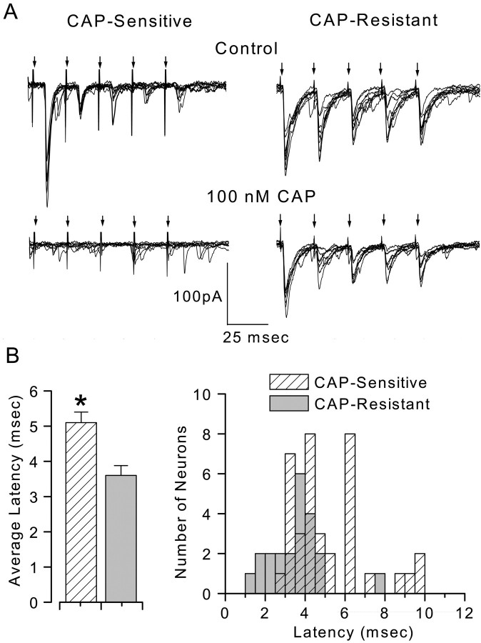
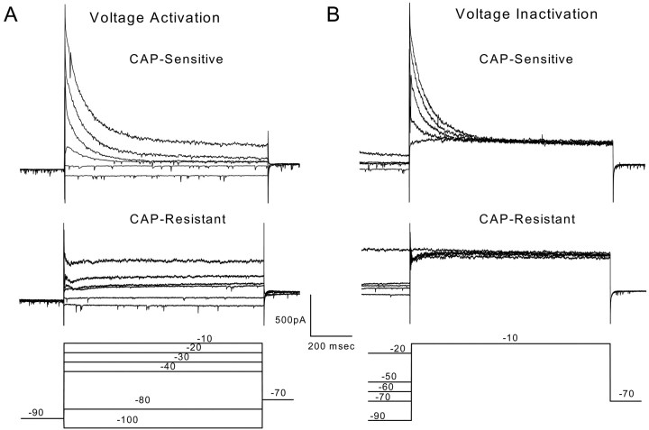
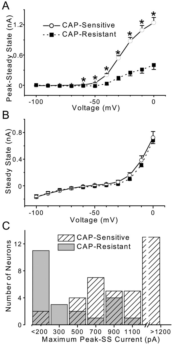
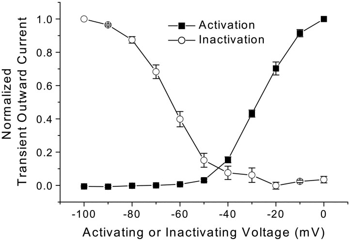
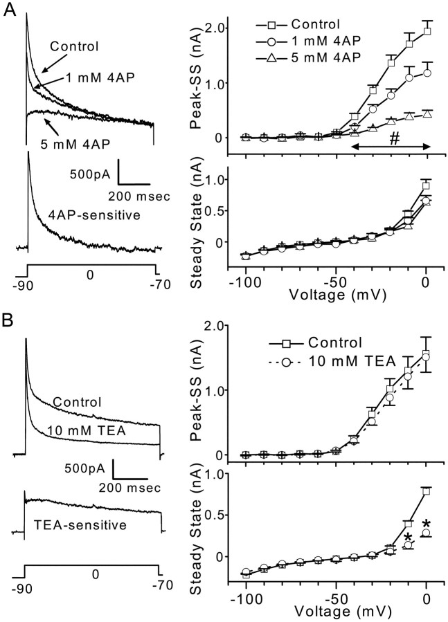
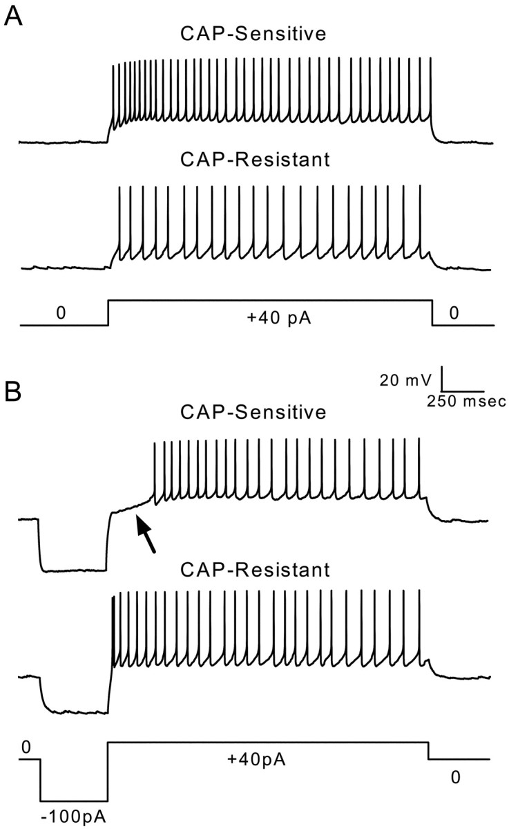
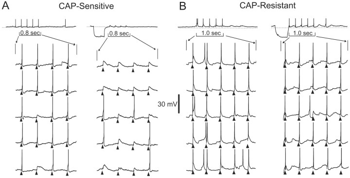
Cranial visceral afferents innervate second-order nucleus tractus solitarius (NTS) neurons via myelinated (A-type) and unmyelinated (C-type) axons in the solitary tract (ST). A- and C-type afferents often evoke reflexes with distinct performance differences, especially with regard to their frequency-dependent properties. In horizontal brainstem slices, we used the vanilloid receptor 1 agonist capsaicin (CAP; 100 nm) to identify CAP-sensitive and CAP-resistant ST afferent pathways to second-order NTS neurons and tested whether these two groups of neurons had similar intrinsic potassium currents. ST stimulation evoked monosynaptic EPSCs identified by minimal synaptic jitter (<150 microsec) and divided into two groups: CAP-sensitive (n = 37) and CAP-resistant (n = 22). EPSCs in CAP-sensitive neurons had longer latencies (5.1 +/- 0.3 vs 3.6 +/- 0.3 msec; p = 0.001) but similar jitter (p = 0.57) compared with CAP-resistant neurons, respectively. Transient outward currents (TOCs) were significantly greater in CAP-sensitive than in CAP-resistant neurons. Steady-state currents were similar in both groups. 4-Aminopyridine or depolarized conditioning blocked the TOC, but tetraethylammonium had no effect. Voltage-dependent activation and inactivation of TOC were consistent with an A-type K+ current, I(KA). In current clamp, the activation of I(KA) reduced neuronal excitability and action potential responses to ST transmission. Our results suggest that the potassium-channel differences of second-order NTS neurons contribute to the differential processing of A- and C-type cranial visceral afferents beginning as early as this first central neuron. I(KA) can act as a frequency transmission filter and may represent a key target for the modulation of temporal processing of reflex responsiveness such as within the baroreflex arc.


Figures

Fig. 1.
Fig. 1.
CAP sensitivity of ST-evoked synaptic responses in mNTS neurons. ST stimulation [five shocks (arrows) at 20 msec intervals] consistently triggered EPSCs in these mNTS neurons. Neurons with synaptic jitters (SD of latency) of <150 μsec were selected for study and considered to be second-order mNTS neurons. The holding potential was −70 mV. The sustained application of CAP (100 nm) blocked ST-evoked EPSCs in CAP-sensitive neurons but not in the remaining neurons (CAP-resistant). A,Left, Nine successive ST-evoked EPSCs from one such CAP-sensitive neuron before (top) and 5 min after (bottom) CAP application. A,Right, ST EPSCs from a representative CAP-resistant neuron before (top) and 5 min after (bottom) CAP application. B,Left, Average latency of ST-evoked EPSCs in CAP-sensitive neurons (hatched bar;n = 37) was significantly longer than in CAP-resistant neurons (solid bar; n= 22; *p = 0.0016; Student'st test). B, Right, Histogram (bin size, 0.5 msec) of the distribution for these individual latencies in CAP-resistant (solid bars) and CAP-sensitive (hatched bars) neurons. Although group average latencies were different, the two overlapped considerably.
Fig. 2.
Fig. 2.
Voltage-dependent activation and inactivation of outward currents in two representative second-order mNTS neurons, one CAP-sensitive and one CAP-resistant. A, Prolonged depolarization evoked prominent TOCs in CAP-sensitive neurons but not in CAP-resistant neurons. All neurons were held at −70 mV before each test sequence. Neurons were preconditioned at −90 mV for 500 msec followed by step commands to test potentials (−100 to 0 mV in 10 mV increments). For clarity, only six representative current responses (A, bottom) are displayed. The large outward current to sustained depolarization of the CAP-sensitive neuron (top) rapidly decayed. Current traces from a representative CAP-resistant neuron (middle) show little such TOC. B, Conditioning neurons at depolarized potentials suppressed the TOC component in the CAP-sensitive neuron (top) but had little effect on the outward currents of CAP-resistant (middle) neurons (representative neurons). Whole-cell currents were measured in response to step voltage commands. Between sweeps, neurons were held at −70 mV. Conditioning steps varied in 10 mV increments from −100 to 0 mV. Conditioning was followed by a prolonged (1200 msec) step to −10 mV. For clarity, only 5 of the 11 sweeps are shown. Voltage step protocols are outlined in thebottom panels.
Fig. 3.
Fig. 3.
Summary of average outward currents in CAP-sensitive (n = 37) and CAP-resistant (n = 22) mNTS neurons. Voltage-dependent TOCs (Peak − SS) and SSCs. Voltage step protocols were identical to those in Figure 2. Points are averages ± SEM. A, CAP-sensitive neurons (solid line) had a significantly larger TOC compared with CAP-resistant neurons (dashed line; p < 0.0001; RM ANOVA). Comparison of the TOC observed at specific voltages revealed that TOCs were larger in CAP-sensitive neurons beginning at −60 mV and extending to 0 mV (one-way ANOVA; *p < 0.001). B, Average SSCs were similar (p = 0.752; RM ANOVA) in CAP-sensitive (solid line) and CAP-resistant (dashed line) neurons. C, Distribution of peak TOCs in CAP-sensitive and CAP-resistant mNTS second-order neurons. CAP-sensitive neurons (hatched bars) generally expressed larger TOCs than did CAP-resistant neurons (solid bars), although there is some overlap.
Fig. 4.
Fig. 4.
Summary of mean activation and inactivation relationships for TOCs of CAP-sensitive neurons. Voltage protocols were identical to those in Figure 2. Average normalized peak TOCs ± SEM (n = 37) are plotted against activation voltage (▪) or conditioning voltage (○). These voltage-dependent activation and inactivation characteristics of TOCs resemble those reported for A-type, 4-AP-sensitive potassium currents.
Fig. 5.
Fig. 5.
Pharmacological sensitivity of outward currents in CAP-sensitive mNTS neurons. TOC was calculated as the peak − SSC and SSC measured at the end of the test step. Command voltage step protocols are indicated in the bottom traces ofA and B. Average values are means ± SEM in each summary relationship. A,Left, Three current traces from a representative experiment generated by the step command in the bottom trace show the effects of 1 and 5 mm 4-AP on TOCs and SSCs. 4-AP reduced the TOC in this neuron in a dose-dependent manner but did not affect the SSCs. The middle trace is a difference current obtained by subtracting the control and 5 mm 4-AP traces to yield the net 4-AP sensitive current.Right, Summary data for 1 and 5 mm 4-AP on TOCs and SSCs in five CAP-sensitive neurons. Points are averages ± SEM in controls (■), with 1 mm 4-AP (○), and 5 mm 4-AP (▵). Both 1 and 5 mm4-AP caused a significant reduction in voltage-activated TOCs compared with controls (p = 0.0001; RM ANOVA). Both 1 and 5 mm 4-AP reduced the TOCs observed at −40 through 0 mV (#p < 0.025; one-way ANOVA and Fisher's PLSD). SSCs were not affected by 4-AP (bottom right; p = 0.262; RM ANOVA). These results suggest that the TOC observed in CAP-sensitive mNTS neurons is an A-type potassium current. B, Left, Two representative current traces evoked by steps to zero potential from −90 mV conditioning levels. The middle trace is a difference current obtained by subtracting the control and 10 mm TEA traces to yield the net TEA-sensitive current.Right, Summary data for TEA (10 mm) in seven CAP-sensitive neurons. TEA had no effect on the TOC (p = 0.6626; RM ANOVA) but reduced the SSCs (p = 0.0014; RM ANOVA) at −10 and 0 mV (*p < 0.0007; one-way ANOVA with Fisher's PLSD).
Fig. 6.
Fig. 6.
The differential expression of A-type potassium currents produces different firing properties in second-order CAP-sensitive and CAP-resistant mNTS neurons. A, Representative voltage traces measured in current-clamp mode from a CAP-sensitive (toptrace) and a CAP-resistant (middle trace) neuron show evoked firing produced by current injection shown in the current command below (+40 pA). Both neurons fire repeated action potentials with little or no delay after the onset of current injection. B, Representative voltage traces measured in current-clamp mode from the same CAP-sensitive (top trace) and CAP-resistant (middle trace) neurons in A showing evoked firing after negative current-evoked membrane hyperpolarization and subsequent depolarization (−100 and +40 pA, step current command shown in bottom trace). The hyperpolarization caused a delay in the production of current-evoked action potentials (arrow) in the CAP-sensitive neuron. In contrast, there was no delay in firing produced by hyperpolarization in the CAP-resistant neurons. The CAP-sensitive neuron from which these voltage traces were recorded displayed a largeIKA-like current. The CAP-resistant neuron did not contain such a current. The delay in evoked firing was likely caused by hyperpolarization-induced removal of inactivation of A-type potassium currents.
Fig. 7.
Fig. 7.
A-type potassium currents have an impact on the processing of ST-evoked responses in second-order mNTS neurons.A, Representative sequential voltage traces measured in current-clamp mode from a representative CAP-sensitive neuron (same neuron as Fig. 6) showing ST-evoked membrane potential responses.Top traces show a complete, 2.5 sec test with ST-evoked EPSPs and action potentials. Bottom traces show expanded sweeps of five consecutive tests to better display responses to bursts of five ST shocks (triangles). Note that the voltage scaling of the expanded sweeps truncates action potentials. ST stimulation in controls produced EPSPs that reliably generated action potentials (A, left). Only 2 of 20 ST shocks failed to trigger an action potential in this example. Introduction of a hyperpolarizing current step (top,long trace) suppressed action potentials but reliably produced EPSPs to ST shocks in this CAP-sensitive neuron (A, right). B, Tests similar to those shown in A were conducted on a representative CAP-resistant neuron (same neuron as Fig. 6). ST stimulation (triangles) reliably produced EPSPs that generated action potentials with and without conditioning hyperpolarization. These observations suggest that the expression of A-type currents has a differential impact on the processing of afferent inputs in CAP-sensitive and CAP-resistant second-order mNTS neurons.

References

    1. Andresen MC, Kunze DL. Nucleus tractus solitarius: gateway to neural circulatory control. Annu Rev Physiol. 1994;56:93–116. - PubMed
    1. Andresen MC, Mendelowitz D. Sensory afferent neurotransmission in caudal nucleus tractus solitarius: common denominators. Chem Senses. 1996;21:387–395. - PubMed
    1. Andresen MC, Yang M. Non-NMDA receptors mediate sensory afferent synaptic transmission in medial nucleus tractus solitarius. Am J Physiol. 1990;259:H1307–H1311. - PubMed
    1. Bevan S, Szolcsányi J. Sensory neuron-specific actions of capsaicin: mechanisms and applications. Trends Pharmacol Sci. 1990;11:330–333. - PubMed
    1. Bevan S, Hothi S, Hughes G, James IF, Rang HP, Shah K, Walpole CS, Yeats JC. Capsazepine: a competitive antagonist of the sensory neurone excitant capsaicin. Br J Pharmacol. 1992;107:544–552. - PMC - PubMed

Publication types

MeSH terms

LinkOut - more resources