. 2002 Oct 1;99(20):12825-30.
doi: 10.1073/pnas.202474099. Epub 2002 Sep 11.

Hypoxia and acidosis activate cardiac myocyte death through the Bcl-2 family protein BNIP3

Affiliations

Hypoxia and acidosis activate cardiac myocyte death through the Bcl-2 family protein BNIP3

Lori A Kubasiak et al. Proc Natl Acad Sci U S A. .

Abstract

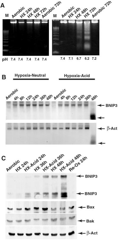
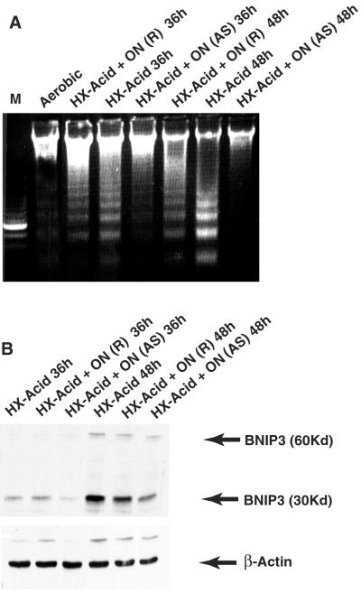
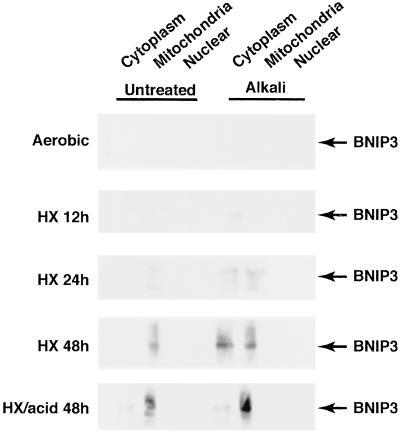
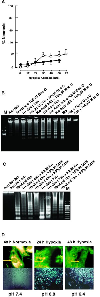
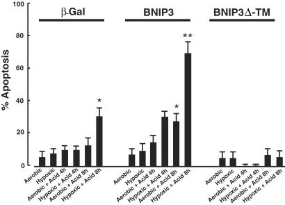
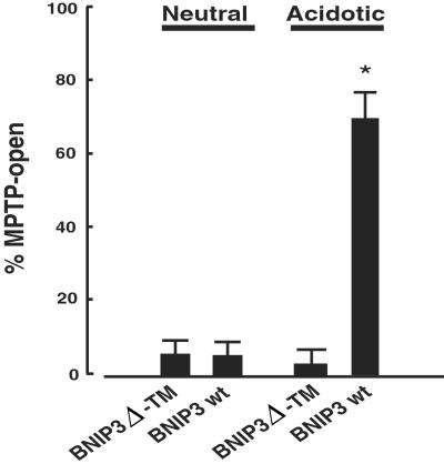
Coronary artery disease leads to injury and loss of myocardial tissue by deprivation of blood flow (ischemia) and is a major underlying cause of heart failure. Prolonged ischemia causes necrosis and apoptosis of cardiac myocytes and vascular cells; however, the mechanisms of ischemia-mediated cell death are poorly understood. Ischemia is associated with both hypoxia and acidosis due to increased glycolysis and lactic acid production. We recently reported that hypoxia does not induce cardiac myocyte apoptosis in the absence of acidosis. We now report that hypoxia-acidosis-associated cell death is mediated by BNIP3, a member of the Bcl-2 family of apoptosis-regulating proteins. Chronic hypoxia induced the expression and accumulation of BNIP3 mRNA and protein in cardiac myocytes, but acidosis was required to activate the death pathway. Acidosis stabilized BNIP3 protein and increased the association with mitochondria. Cell death by hypoxia-acidosis was blocked by pretreatment with antisense BNIP3 oligonucleotides. The pathway included extensive DNA fragmentation and opening of the mitochondrial permeability transition pore, but no apparent caspase activation. Overexpression of wild-type BNIP3, but not a translocation-defective mutant, activated cardiac myocyte death only when the myocytes were acidic. This pathway may figure significantly in muscle loss during myocardial ischemia.


Figures

Figure 1
Figure 1
BNIP3 induction correlates with increased apoptosis. (A) Cardiac myocytes were subjected to hypoxia with (Left) or without (Right) medium change, harvested at the indicated times, and processed for genomic fragmentation assays. The pH of the media at the time of harvesting is shown at the bottom. (B) Northern blots of cardiac myocyte RNA extracted from hypoxic cultures. (C) Western blot analysis of proteins from hypoxic cardiac myocytes as in A. Anti-BNIP3 recognizes two bands at approximately 60 and 30 kDa, corresponding to SDS-resistant homodimers and monomers, respectively. Lower gels show the same blot probed with anti-Bax, Bak, and β-actin. Results are representative of at least three experiments.
Figure 2
Figure 2
BNIP3 antisense inhibits programmed death of cardiac myocytes. (A) Cultures were incubated with BNIP3 (AS) or random sequence (R) oligonucleotides (ON) as described in Materials and Methods, subjected to hypoxia-acidosis as indicated, and analyzed for DNA fragmentation. (B) Cardiac myocytes were treated with oligonucleotides as in A, and extracted proteins were analyzed by Western blots with anti-BNIP3 and β-actin. Results are representative of three experiments.
Figure 3
Figure 3
Association of BNIP3 with subcellular fractions. Cardiac myocytes were subjected to hypoxia as described in Fig. 1. At the indicated times cells were harvested, rinsed, lysed, and subjected to alkaline solubilization (right-hand gels) as described in Materials and Methods. After treatments, samples were separated into subcellular fractions and analyzed by Western blots. Blots were reprobed with antisuccinate dehydrogenase probes to define the purity of fractions (not shown). Results are representative of three separate experiments.
Figure 4
Figure 4
Characteristics of programmed cell death by BNIP3. (A) Cardiac myocytes were subjected to hypoxia-acidosis as described in Fig. 1. At the indicated times, samples of media were taken for analysis of LDH activity (open circles) or plates were stained with trypan blue (closed circles). Data are expressed as percent of cells stained with trypan blue or percent LDH released relative to total LDH in homogenates. (B) Cardiac myocytes were subjected to hypoxia-acidosis in the absence or presence of the broad-range caspase inhibitor Boc-D, as indicated. Staurosporine (Sta; 1.0 μM for 8 h) is shown as a positive control. (C) Cardiac myocytes were subjected to hypoxia-acidosis in the absence or presence of the MPTP inhibitors BA or DUB, as indicated. (D) Cardiac myocytes were exposed to normoxic or hypoxia-acidosis conditions. At the times indicated, cells were loaded with MitoTracker Red dye and analyzed by confocal microscopy. Arrows indicate intense staining around nuclei in aerobic myocytes and reduced/diffuse staining under hypoxia. Results are representative of three experiments.
Figure 5
Figure 5
Programmed death of BNIP3-transfected cardiac myocytes. Cardiac myocytes were transfected with expression plasmids containing β-Gal plus empty vector, β-Gal with BNIP3, or β-Gal with BNIP3 Δ-TM, as indicated. After 48 h, transfected cultures were exposed to continued normoxic culture or to hypoxia, acidosis, or hypoxia + acidosis as described in Materials and Methods. At the indicated times, plates were rinsed, costained with 5-bromo-4-chloro-3-indolyl β-D-galactoside and Hoechst 33342, and visualized by microscopy. Bars indicate SEM from at least 200 5-bromo-4-chloro-3-indolyl β-D-galactoside-positive cells per condition. *, P < 0.02, and **, P < 0.01, refer to the respective condition compared with aerobic controls. BNIP3 Δ-TM transfection with 8-h hypoxia + acid was significantly different from either β-Gal (P < 0.05) or BNIP3 8-h hypoxia + acid (P < 0.01).
Figure 6
Figure 6
Acid-mediated opening of MPTP in BNIP3-transfected myocytes. Cardiac myocytes were cotransfected with pGFP and BNIP3 or BNIP3 Δ-TM, as indicated. After 48 h, transfected cultures were exposed to continued culture at neutral pH or to acidosis for an additional 8 h as described in Materials and Methods. Cultures were stained with MitoTracker Red dye, fixed, and analyzed by microscopy. At least 50 GFP-positive myocytes per condition were scored from four separate dishes.

References

    1. Jennings R B, Steebergen C, Reimer K A. Monogr Pathol. 1995;37:47–80. - PubMed
    1. Vanoverschelde J-L J, Wijns W, Borgers M, Heyndrickx G, Depre C, Flambeng W, Melin J A. Circulation. 1997;95:1961–1971. - PubMed
    1. Zurbier C J, van Iterson M, Ince C. Cardiovasc Res. 1999;44(3):488–497. - PubMed
    1. Narula J, Hajjar R J, Dec G W. Cardiol Clin. 1998;16(4):691–710. - PubMed
    1. Dennis S C, Gevers W, Opie L H. J Mol Cell Cardiol. 1991;23:1077–1086. - PubMed

Publication types

MeSH terms

LinkOut - more resources