Review
. 2002 Sep;130(1):22-46.
doi: 10.1104/pp.004770.

Inositol phospholipid metabolism in Arabidopsis. Characterized and putative isoforms of inositol phospholipid kinase and phosphoinositide-specific phospholipase C

Affiliations
Review

Inositol phospholipid metabolism in Arabidopsis. Characterized and putative isoforms of inositol phospholipid kinase and phosphoinositide-specific phospholipase C

Bernd Mueller-Roeber et al. Plant Physiol. 2002 Sep.

Abstract

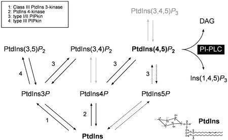
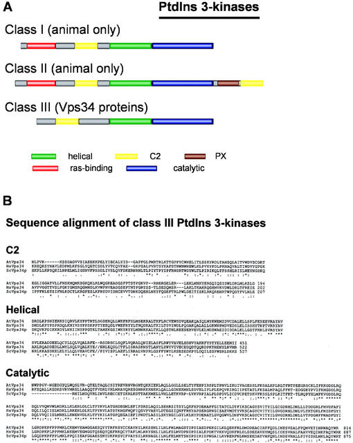
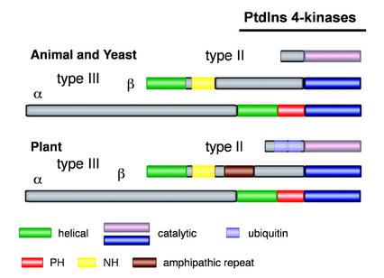
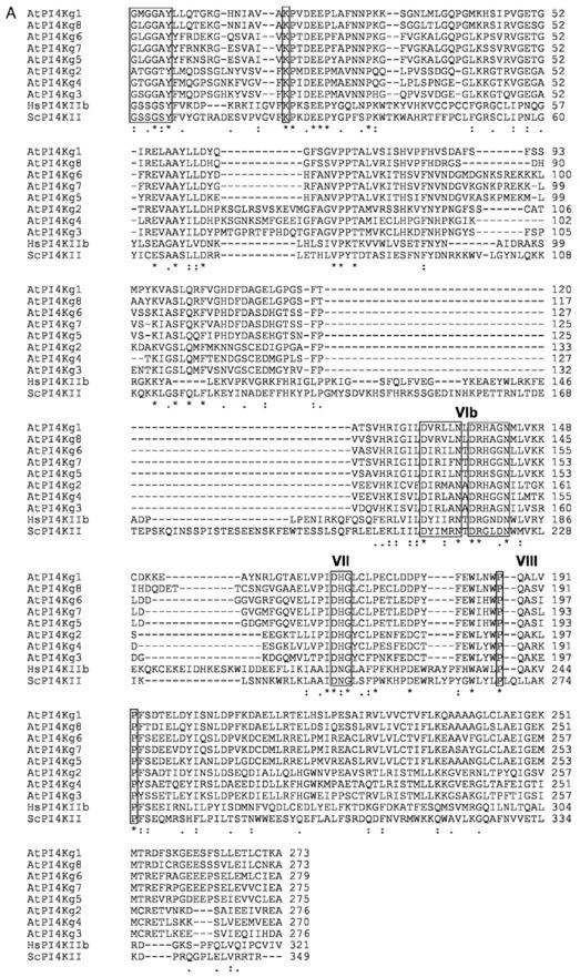
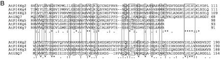
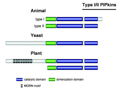
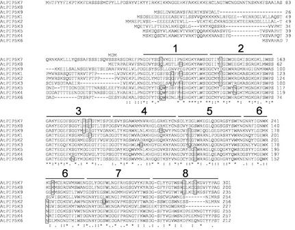
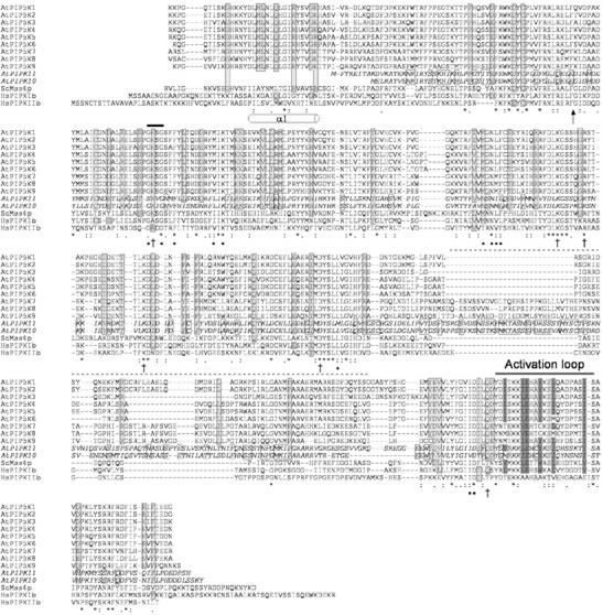
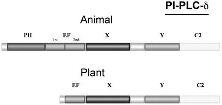
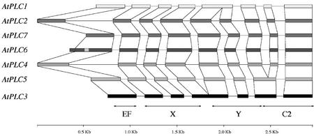
Phosphoinositides (PIs) constitute a minor fraction of total cellular lipids in all eukaryotic cells. They fulfill many important functions through interaction with a wide range of cellular proteins. Members of distinct inositol lipid kinase families catalyze the synthesis of these phospholipids from phosphatidylinositol. The hydrolysis of PIs involves phosphatases and isoforms of PI-specific phospholipase C. Although our knowledge of the roles played by plant PIs is clearly limited at present, there is no doubt that they are involved in many physiological processes during plant growth and development. In this review, we concentrate on inositol lipid-metabolizing enzymes from the model plant Arabidopsis for which biochemical characterization data are available, namely the inositol lipid kinases and PI-specific phospholipase Cs. The biochemical properties and structure of characterized and genome-predicted isoforms are presented and compared with those of the animal enzymes to show that the plant enzymes have some features clearly unique to this kingdom.


Figures

Figure 1
Figure 1
PI metabolism. The different steps in the synthesis of PIs and the lipid kinases catalyzing the different reactions are indicated. PtdIns(3,4,5)P3 is present in animal cells but has not been detected in plant tissues, so far. In animal cells, PtdIns(3,4)P2 can be generated from PtdIns4P by a PtdIns 3-kinase or by an as-yet-unidentified PIPkin from PtdIns3P. Plant cells do not contain any homolog of the heterodimeric inositol lipid 3-kinases that are able to phosphorylate PtdIns4P to PtdIns(3,4)P2 and PtdIns(4,5)P2 to PtdIns(3,4,5)P3. PtdIns(4,5)P2 can be synthesized by type I and type II PIPkins from PtdIns4P and PtdIns5P, respectively. On the basis of sequence comparison, plants cells do not possess type II PIPkins. PtdIns5P is present in plants, but an enzyme capable of producing it has not been identified.
Figure 2
Figure 2
Domain structure representation of PtdIns 3-kinases. A, The domains conserved in the catalytic subunits of the three PtdIns 3-kinase classes are represented by colored boxes. B, Vps34 proteins from Brewer's yeast (Vps34p; accession no. X53531), Arabidopsis (AtVps34; accession no. U10669), and human (HsVps34; accession no. Z46973) were aligned using the ClustalW program from the MacVector package. Identical, conserved, and semiconserved residues are indicated below the alignment by *, :, and ., respectively. // indicates a large gap in the sequence.
Figure 3
Figure 3
Domain structure representation of animal, yeast, and plant PtdIns 4-kinases. The various conserved domains are represented by colored boxes. The catalytic domains of type II and III PtdIns 4-kinase share no homology and are therefore shown in different colors. Some plant type II PtdIns 4-kinases contain one or two ubiquitin-like domains.
Figure 4
Figure 4
Comparison of putative Arabidopsis type II PtdIns 4-kinases with the catalytic domain of type II PtdIns 4-kinases from human and Brewer's yeast (A) and with ubiquitin (B). Sequences were aligned with the ClustalW program and adjusted by hand. Identical, conserved, and semiconserved residues are indicated below the alignment by *, :, and ., respectively. A, Alignment of predicted catalytic domains. The location of candidate kinase subdomains I, II, VIb, and VII, identified by Minogue et al. (2001), are indicated in roman numerals above the alignment. A candidate subdomain VIII is also indicated. The kinase motifs in subdomains I, II, VIb, and VII identified by Minogue et al. (2001) are boxed. The two P residues in the PXXXXP motif in the candidate subdomain VIII are also boxed. B, Alignment of the N-terminal domains of six of the Arabidopsis putative type II PtdIns 4-kinases with ubiquitin 7 from Arabidopsis (AtUBQ7; accession no. NM_129118). AtUBQ7, AtPI4Kγ2, AtPI4Kγ3, and AtPI4Kγ4 contain two ubiquitin domains, whereas AtPI4Kγ5, AtPI4Kγ6, and AtPI4Kγ7 contain only one. Amino acid residues identical or conserved at identical positions in the two ubiquitin domains of AtUBQ7 and the ubiquitin-like domains of Arabidopsis type II PtdIns 4-kinases are boxed.
Figure 4
Figure 4
Comparison of putative Arabidopsis type II PtdIns 4-kinases with the catalytic domain of type II PtdIns 4-kinases from human and Brewer's yeast (A) and with ubiquitin (B). Sequences were aligned with the ClustalW program and adjusted by hand. Identical, conserved, and semiconserved residues are indicated below the alignment by *, :, and ., respectively. A, Alignment of predicted catalytic domains. The location of candidate kinase subdomains I, II, VIb, and VII, identified by Minogue et al. (2001), are indicated in roman numerals above the alignment. A candidate subdomain VIII is also indicated. The kinase motifs in subdomains I, II, VIb, and VII identified by Minogue et al. (2001) are boxed. The two P residues in the PXXXXP motif in the candidate subdomain VIII are also boxed. B, Alignment of the N-terminal domains of six of the Arabidopsis putative type II PtdIns 4-kinases with ubiquitin 7 from Arabidopsis (AtUBQ7; accession no. NM_129118). AtUBQ7, AtPI4Kγ2, AtPI4Kγ3, and AtPI4Kγ4 contain two ubiquitin domains, whereas AtPI4Kγ5, AtPI4Kγ6, and AtPI4Kγ7 contain only one. Amino acid residues identical or conserved at identical positions in the two ubiquitin domains of AtUBQ7 and the ubiquitin-like domains of Arabidopsis type II PtdIns 4-kinases are boxed.
Figure 5
Figure 5
Domain structure representation of animal, yeast, and plant type I/II PIPkins. The conserved domains are indicated by boxes of different colors.
Figure 6
Figure 6
Alignment of the N-terminal MORN domain of type I/II PIPkins from Arabidopsis. The alignment was generated with the ClustalW program and adjusted by hand. Identical, conserved, and semiconserved residues are indicated below the alignment by *, :, and ., respectively. The eight MORN motifs (consensus sequence: Y-Q/E-G-E/Q-T-X-N-G-K-X-H-G-Y-G) are indicated by black lines on top of the alignment. Residues within the MORN consensus sequence that are conserved in only a few of the sequences are boxed.
Figure 7
Figure 7
Comparison of the catalytic domain of type I and II PIPkins from animal, yeast, and Arabidopsis. The PIPkin catalytic domain of the nine Arabidopsis PIPkins, Brewer's yeast Mss4p and human PIPKIβ and PIPKIIβ were aligned using the ClustalW program. Arabidopsis subfamily A sequences are in italics. Residues identical in all sequences are indicated by *. The residues conserved or semiconserved are marked with : and ., respectively. Residues identical or conserved in Arabidopsis sequences only are boxed and have gray backgrounds. Residues identical in AtPIPK10 and AtPIPK11 only, and not conserved in other proteins, are boxed. The activation loop is indicated. Residues in HsPIPKIIb proposed to interact with ATP and PtdIns5P are indicated with ● and †, respectively (Rao et al., 1998). Residues in the activation loop that are conserved among type I or II PIPkins are shaded in dark and light gray, respectively, only when they are also conserved in Arabidopsis sequences. The position of the first α-helix of the catalytic domain of human PIPKIIβ (Rao et al., 1998) is shown under the sequence alignment, and the location of the end of the N-terminal dimerization domain is indicated by an arrow. The inserts in the catalytic domains are marked by a dotted line on top of the alignment. The GXXG motif conserved in the catalytic domain of protein kinases and type II PtdIns 4-kinases is indicated by a black bar over the alignment.
Figure 8
Figure 8
A phylogenic tree of subfamily B AtPIPkins. Full-length protein sequences were aligned and the tree was constructed using the PAUP software (Phylogenetic Analysis Using Parsimony, version 4.0b4, Sinauer Associates, Sunderland, MA) from 12,000 replicates. An identical tree is obtained if the sequences of the catalytic domains alone are compared. The three pairs of duplicated genes are circled, and the duplicated blocks they belong to are indicated (Vision et al., 2000).
Figure 9
Figure 9
Schematic representation of the genes encoding characterized and putative type I/II PIPkins from Arabidopsis. The introns and exons for AtPIP5K1, AtPIP5K5, AtPIP5K7, and AtPIP5K8 were determined by comparing the mRNA and gene sequences, whereas for the other genes, they were deduced by comparison with the four known gene structures and examination of the exon-intron splice junctions. The regions coding for the different conserved domains are indicated by double arrows. Dim, Dimerization domain.
Figure 10
Figure 10
Schematic representation of Fab1 proteins. The conserved domains are represented by blocks of different colors. The intervals between the conserved domains vary among species. Two of the four Arabidopsis genes encoding putative Fab1 proteins do no contain a FYVE domain.
Figure 11
Figure 11
Alignment of the seven PI-PLCs from Arabidopsis. The accession numbers for the different proteins are given in Table II. The sequences were aligned using the ClustalW program. The four conserved domains are indicated. The X and Y domains together form the catalytic domain of the enzymes. Because no mRNA or EST sequences for AtPLC6 have been obtained, the sequence shown here is the predicted one that fits best with the other sequences.
Figure 12
Figure 12
Representation of the modular domain arrangements of PI-PLC δ isozymes from animals and plants. The conserved domains are represented by blocks of different colors. The EF-hand domain of plant PI-PLCs corresponds to the second loop of the EF-hand domain of animal PI-PLCs. The X and Y domains constitute together the catalytic domain of PI-PLCs.
Figure 13
Figure 13
Schematic representation of the seven PI-PLC genes from Arabidopsis. The introns and exons for AtPLC1, AtPLC2, AtPLC3, AtPLC4, and AtPLC5 were determined by comparing the mRNA and gene sequences, whereas for the other three genes, they were deduced by comparison with AtPLC1-5 and examination of the exon-intron splice junctions. In AtPLC6, two putative start codons can be identified. In addition, an insert in the first exon of this gene is present, and indicated by a gray box. The regions coding for the different conserved domains are indicated by double arrows.

References

    1. Alexandre J, Lassalles JF, Kado RD. Opening of Ca2+channels in isolated red beet vacuole membrane by inositol 1,4,5-trisphosphate. Nature. 1990;343:567–570.
    1. Ananthanarayanan B, Das S, Rhee SG, Murray D, Cho W. Membrane targeting of C2 domains of phospholipase C-δ isoforms. J Biol Chem. 2001;277:3568–3575. - PubMed
    1. Audhya A, Foti M, Emr SD. Distinct roles for the yeast phosphatidylinositol 4-kinases, Stt4p and Pik1p, in secretion, cell growth, and organelle membrane dynamics. Mol Biol Cell. 2000;11:2673–2689. - PMC - PubMed
    1. Bae YS, Cantley LG, Chen CS, Kim SR, Kwon KS, Rhee SG. Activation of phospholipase C-γ by phosphatidylinositol 3,4,5-trisphosphate. J Biol Chem. 1998;273:4465–4469. - PubMed
    1. Balla T. Phosphatidylinositol 4-kinases. Biochim Biophys Acta. 1998;1436:69–85. - PubMed

MeSH terms

Substances