. 2002 Sep;130(1):90-101.
doi: 10.1104/pp.006288.

Molecular identification of cytosolic, patatin-related phospholipases A from Arabidopsis with potential functions in plant signal transduction

Affiliations

Molecular identification of cytosolic, patatin-related phospholipases A from Arabidopsis with potential functions in plant signal transduction

André Holk et al. Plant Physiol. 2002 Sep.

Abstract

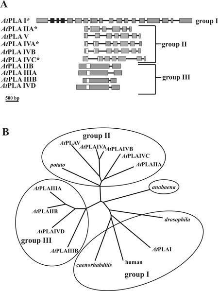
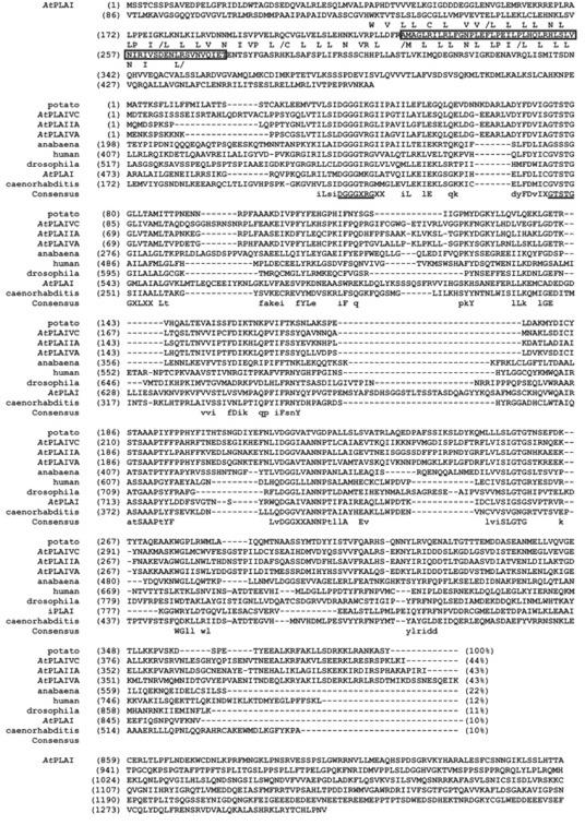
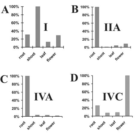
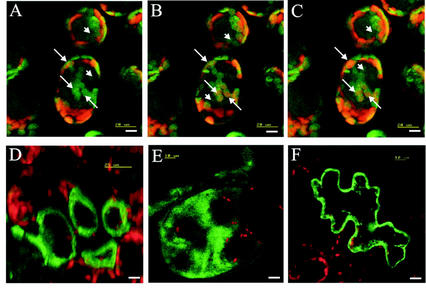
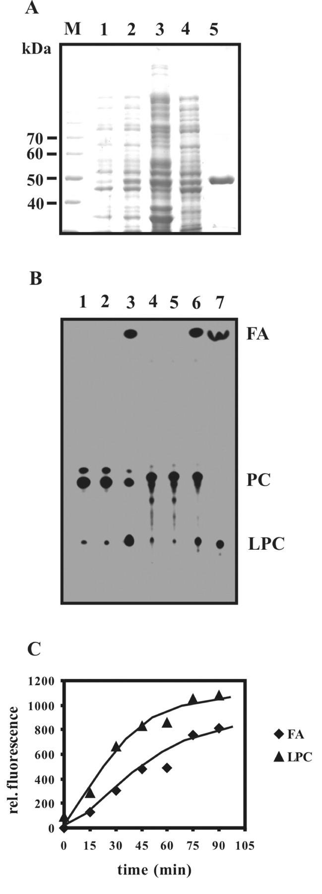
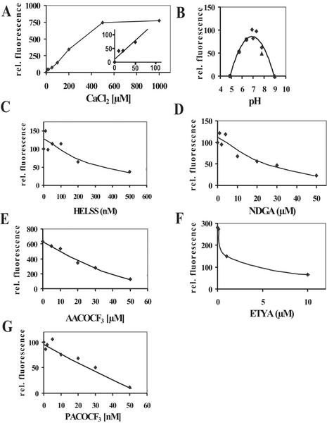
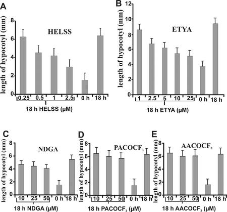
Rapid activation of phospholipase A (PLA) by auxin or plant-pathogen interaction suggests a function in signal transduction for this enzyme, but the molecular identification of a cytosolic PLA carrying out this function remains open. We isolated four cDNA sequences from Arabidopsis (ecotype Columbia), AtPLA I, AtPLA IIA, AtPLA IVA, and AtPLA IVC, which are members of the patatin-related PLA gene family in plants and which are homologous to the animal Ca(2+)-independent PLA(2) gene family. Expression was measured by reverse transcriptase-polymerase chain reaction, and AtPLA I transcripts were found preferentially in shoots, AtPLA IIA and AtPLA IVA in roots, and AtPLA IVC in flowers. Transient expression of the four PLA-green fluorescent protein fusion proteins in tobacco (Nicotiana tabacum) leaves showed they were located in the cytosol and not in the vacuoles. Surprisingly, AtPLA::green fluorescent protein was also localized to chloroplasts. The enzymatic activity of the purified recombinant AtPLA IVA toward phosphatidylcholine was dependent on Ca(2+), saturated at 0.5 mM, and had a pH optimum of about 7.0. It had both PLA(1) and PLA(2) specificity. The enzyme showed in vitro highest sensitivity toward the PLA(2) inhibitors palmitoyltrifluoromethyl ketone (PACOCF(3), K(i) approximately 30 nM), arachidonyltrifluoromethyl ketone (AACOCF(3), K(i) approximately 25 microM), and tetrahydro-3-(1-naphtalenyl)-2H-pyran-2-one (K(i) approximately 200 nM) and was also sensitive to other previously used inhibitors 5,8,11,14-eicosatetraynoic acid (K(i) approximately 3 microM) and nordihydroguajaretic acid (K(i) approximately 15 microM). The influence of these PLA(2) inhibitors on elongation in etiolated Arabidopsis seedlings was tested, and tetrahydro-3-(1-naphtalenyl)-2H-pyran-2-one and 5,8,11,14-eicosatetraynoic acid inhibited hypocotyl elongation maximally at concentrations close to their K(i) in vitro.


Figures

Figure 1
Figure 1
Gene structure of the patatin-related AtPLA-family in Arabidopsis and a phylogenetic tree of several patatin-related plant PLA and animal iPLA2 sequences. A, Gene structure of the AtPLA family in Arabidopsis. Exons are symbolized by gray boxes, those containing the LRR by black boxes, the catalytic center by white boxes, and introns by black lines. cDNA sequences described in this work are marked by an asterisk. The sequences are deposited in database as follows: AtPLA I (accession no. AC004392), AtPLA IIA (accession no. AC002505), AtPLA IIB (accession no. AC004697), AtPLA IIIA (accession no. AL049655.1), AtPLA IIIB (accession no. AL138648), AtPLA IVA (db_xref GI:4006869), AtPLA IVB (db_xref GI:4006870), AtPLA IVC (db_xref GI:4006871), AtPLA IVD (accession no. AL050352.1), and AtPLA V (accession no. AB016875.1). A 500-bp size standard is indicated. B, Phylogenetic tree produced by the program ClustalW (http://www2.ebi.ac.uk/clustalw/) on the amino acid structures of the AtPLA gene-family, a putative bacterial protein from Anabeana sp. (accession no. AJ269505.1; Rouhiainen et al., 2000), a putative protein from the nematode Caenorhabditis elegans (accession no. AC084197.1), a putative protein from the fruitfly (Drosophila melanogaster; accession no. AE003550.2), an iPLA2 from human (accession no. JC7284; Tanaka et al., 2000), and a patatin class I precursor from potato (accession no. P11768; Mignery et al., 1988) displayed by the program TREEVIEW (Page, 1996).
Figure 2
Figure 2
Alignment of predicted amino acid sequences of isolated PLA cDNAs from Arabidopsis and of selected domains of patatin-related PLAs from other organisms. The conserved residues of LRRs of the consensus sequence LXXLXXLXLXXN/CXXL/IP/RXLXXLXX are highlighted by printing the conserved residues below the AtPLA I sequence. The additional exon found by us and derived from the isolated cDNA of PLA I is boxed. The most highly conserved motifs in the consensus sequence DGGGXRG of the catalytic center and the lipase motif GTSTG are underlined. The PLA amino acid sequences from Arabidopsis AtPLA I, AtPLA IIA, AtPLA IVA, and AtPLA IVC are compared with a putative bacterial protein from Anabeana sp. (accession no. AJ269505.1), a putative protein from the nematode C. elegans (accession no. AC084197.1), a putative protein from the fruitfly (accession no. AE003550.2), an iPLA2 from human (accession no. JC7284), and a patatin class I precursor from potato (accession no. P11768) by using the program vector NTI from InforMax. A consensus sequence is indicated below the sequences.
Figure 3
Figure 3
Expression of mRNA of the AtPLA I, AtPLA IIA, AtPLA IVA, and AtPLA IVC gene in organs of Arabidopsis. The total RNA from roots, shoots, leaves, and flowers was analyzed by competitive RT-PCR using gene-specific internal standards for the genes I, IIA, and IVA or by RT-PCR using actin as external standard for the gene IVC; separated on ethidium-bromide gel; and inverted into gray scale for digitization and quantification. Relative amounts of cDNAs (highest value is set to 100%) are shown in A for I, in B for IIA, in C for IVA, and in D for IVC.
Figure 4
Figure 4
Transient expression of PLA-GFP fusion proteins in tobacco leaf cells as visualized by confocal laser scanning microscopy. A through C, Three consecutive optical sections, moving from the epidermal top side of palisade cells toward the spongy parenchyma side containing, expressed PLA-I-GFP fusion protein. Arrows, Localization of PLA-I-GFP fusion protein in an individual chloroplast in consecutive frames. Arrowheads, Localization of PLA-I-GFP fusion protein in the cytosol. D, Single optical section of palisade parenchyma cells expressing the PLA-IIA-GFP fusion protein. E, Summarized image showing the expressed PLA-IVA-GFP-fusion protein calculated from 16 consecutively taken single optical sections (in z-direction) through a large cell. F, Single optical section of an epidermal cell expressing the PLA-IVC-GFP fusion protein. Bar = 10 μm.
Figure 5
Figure 5
Purification and PLA activity of the recombinant AtPLA IVA protein. A, SDS-PAGE of recombinant AtPLA IVA-purification steps. Lane M, Mr size marker; lane 1, E. coli proteins without induction by isopropyl-β-d-thiogalactosid (IPTG); lane 2, E. coli proteins after induction by 0.5 mm IPTG; lane 3, 12,000g pellet after lysis; lane 4, supernatant of soluble proteins (12,000g centrifugation); lane 5, nickel-nitrilotriacetic acid agarose resin-purified recombinant AtPLA IVA protein. B, Thin-layer chromatogram and comparison of the enzymatic products of PLA digestion of 2-(4,4-difluoro-5-methyl-4-bora-3a,4a-diaza-s-indacene-3-dodecanoyl)-1-hexadecanoyl-sn-glycero-3-phosphocholine (in lanes 1–3) and bis-BODIPY-PC (lanes 4–6) by recombinant protein from gene AtPLA IVA. Lane 1, Monolabeled substrate only; lane 2, assay with monolabeled substrate using equivalent amount to lane 3 of control eluate of an empty-vector purification; lane 3, assay with monolabeled substrate using 0.5 μg of IVA purified protein; lane 4, bis-labeled substrate only; lane 5, bis-labeled substrate using an equivalent amount of control eluate from empty-vector purification; lane 6, bis-labeled substrate using 0.5 μg of purified IVA protein in 6 μL; and lane 7, standards for fatty acid and lysophosphatidylcholine. C, PLA activity test. Time-course experiment with recombinant AtPLA IVA and bis-labeled substrate. The amounts of the remaining substrate and the enzymatic products, fatty acid (FA) and lysophosphatidylcholine (LPC), had different apparent molar efficiencies regarding fluorescence emission because of different spot sizes as captured by video photography so that results are expressed on a relative scale.
Figure 6
Figure 6
Catalytic properties of purified recombinant PLA derived from gene AtPLA IVA. A, Ca2+ dependence of PLA activity; inset, highlighting of low Ca2+ concentrations. B, pH dependence of activity; C, inhibition by HELSS; D, inhibition by NDGA; E, inhibition by AACOCF3; F, inhibition by ETYA; and G, inhibition by PACOCF3. Fluorescent fatty acid was quantified on a relative scale.
Figure 7
Figure 7
Influence of PLA2-inhibitors on auxin-induced hypocotyl elongation growth of Arabidopsis. A, Inhibition by HELSS; B, inhibition by ETYA; C, inhibition by NDGA; D, inhibition by PACOCF3; and E, inhibition by AACOCF3. se is shown (25–40 individuals per sample).

References

    1. Ackermann JE, Conde-Frieboes K, Dennis EA. Inhibition of macrophage Ca2+-independent phospholipase A2 by bromoenol lactone and trifluoromethyl ketones. J Biol Chem. 1995;270:445–450. - PubMed
    1. Andrews DL, Beames B, Summers MD, Park WD. Characterization of the lipid acyl hydrolase activity of the major potato (Solanum tuberosum) tuber protein, patatin, by cloning an abundant expression in a baculovirus vector. Biochem J. 1988;252:199–206. - PMC - PubMed
    1. Balsinde J, Balboa MA, Insel PA, Dennis EA. Regulation and inhibition of phospholipase A2. Annu Rev Pharmacol Toxicol. 1999;39:175–189. - PubMed
    1. Balsinde J, Dennis EA. Function and inhibition of intracellular calcium-independent phospholipase A2. J Biol Chem. 1997;272:16069–16072. - PubMed
    1. Benedetti CE, Costa CL, Turcinelli CR, Arruda P. Differential expression of a novel gene in response to coronatine, methyl jasmonate, and wounding in the coi1 mutant of Arabidopsis. Plant Physiol. 1998;116:1037–1042. - PMC - PubMed

Publication types

MeSH terms

LinkOut - more resources