. 2002 Nov 22;277(47):45195-202.
doi: 10.1074/jbc.M205380200. Epub 2002 Sep 12.

Suprabasin, a novel epidermal differentiation marker and potential cornified envelope precursor

Affiliations

Suprabasin, a novel epidermal differentiation marker and potential cornified envelope precursor

Geon Tae Park et al. J Biol Chem. .

Abstract

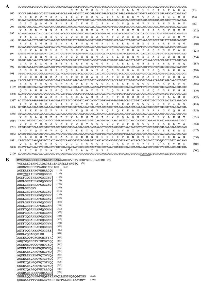
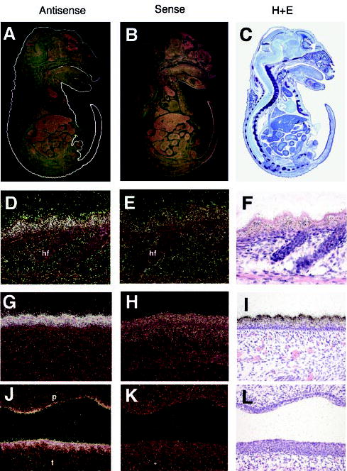
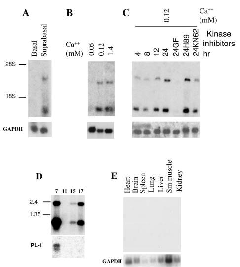
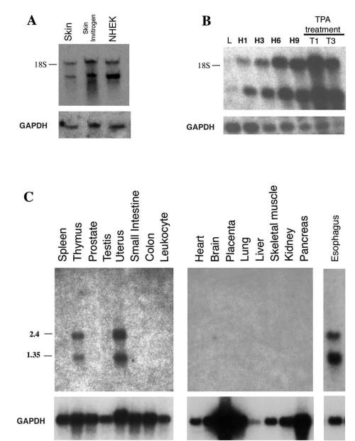
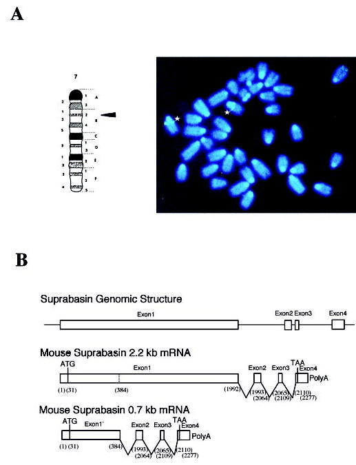
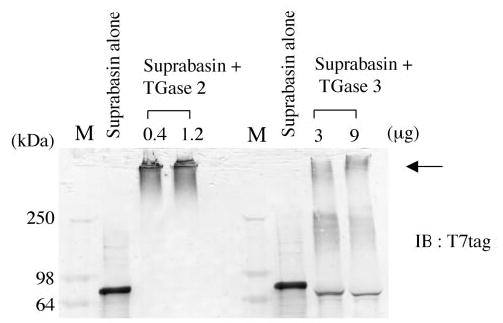
The suprabasin gene is a novel gene expressed in mouse and human differentiating keratinocytes. We identified a partial cDNA encoding suprabasin using a suppression subtractive hybridization method between the proliferative basal and differentiating suprabasal populations of the mouse epidermis. A 3' gene-specific probe hybridized to transcripts of 0.7- and 2.2-kb pairs on Northern blots with specific detection in differentiated keratinocytes of stratified epithelia. The mouse gene was mapped to chromosome 7 by fluorescence in situ hybridization. This region is syntenic to human chromosome band 19q13.1, which contained the only region in the data bases with homology to the mouse suprabasin sequence. During embryonic mouse development, suprabasin mRNA was detected at day 15.5, coinciding with epidermal stratification. Suprabasin was detected in the suprabasal layers of the epithelia in the tongue, stomach, and epidermis. Differentiation of cultured primary epidermal keratinocytes with 0.12 mm Ca(2+) or 12-O-tetradecanoylphorbol-13-acetate treatment resulted in the induction of suprabasin. The 2.2-kb cDNA transcript encodes a protein of 72 kDa with a predicted isoelectric point of 6.85. The translated sequence has an amino-terminal domain, a central domain composed of repeats rich in glycine and alanine, and a carboxyl-terminal domain. The alternatively spliced 0.7-kb transcript encodes a smaller protein that shares the NH(2)- and COOH-terminal regions but lacks the repeat domain region. Cross-linking experiments indicate that suprabasin is a substrate for transglutaminase 2 and 3 activity. Altogether, these results indicate that the suprabasin protein potentially plays a role in the process of epidermal differentiation.


Figures

Fig. 1
Fig. 1. Nucleotide and amino acid sequences of mouse suprabasin
A, the nucleotide sequence of mouse suprabasin cDNA (2277 bp) was aligned with the predicted open reading frame amino acid sequence. Three open triangles indicate the sites of splicing (exon/intron boundaries), and the double-underlined nucleotides indicate the canonical poly(A) addition signal. B, the amino acid sequence of mouse suprabasin has been aligned to show the central repetitive domain region (boxed). The dotted line indicates the different repeat sequence region. The shaded residues highlight a predicted transmembrane region domain. The underlined sequences in the COOH-terminal domain indicate potential protein kinase C phosphorylation sites. Numbers in parentheses at the right side indicate number of residues.
Fig. 2
Fig. 2. Expression of suprabasin
In situ hybridization was performed with antisense (A) and sense (B) suprabasin probes on sagittal sections of E17.5 mouse embryos. C, hematoxylin and eosin (H+E) staining of E17.5 sagittal sections. D, G, and J, 20× magnification of trunk skin (D), tail skin (G), and palate (J) with antisense suprabasin. Corresponding hybridization with sense suprabasin (E, H, and K) and hematoxylin and eosin stain (F, I, and L). hf, hair follicle; p, palate; t, tongue.
Fig. 3
Fig. 3. Northern blot analysis of mouse suprabasin
A, expression of suprabasin in the epidermis of newborn mice. Northern blot of poly(A)+ mRNA (2 μg) from basal and differentiated keratinocytes isolated by discontinuous Percoll gradient from neonatal epidermis. The position of the 18 and 28 S ribosomal RNAs are shown. B, expression of suprabasin in primary mouse keratinocytes cultured and differentiated in vitro by addition of Ca2+. C, time course of suprabasin expression in cultured mouse keratinocytes and after 24 h of treatment with different kinase inhibitors: GF (PKC inhibitor), H89 (PKA inhibitor), or KN62 (CaM kinase II inhibitor). D, mouse embryonic mRNA blot (Clontech). Panel shows 2 μg of poly(A)+ per lane from four embryonic developmental stages (7-, 11-, 15-, and 17-day embryos). RNA size marker is indicated on the left. E, adult multiple tissue mRNA Northern blot (Clontech). Tissue sample is indicated above each lane. All blots were hybridized with 262-bp suprabasin, glyceraldehyde-3-phosphate dehydrogenase, and PL-1 (placenta lactogen 1) probes, as indicated.
Fig. 4
Fig. 4. Northern blot analysis of human suprabasin
A, expression of suprabasin in NHEK. Northern blot of RNA (2 μg) from human skin, commercial preparation of human skin (Invitrogen), and human keratinocytes cultured in vitro in 1.4 mM Ca2+ (NHEK). The position of the 18 S ribosomal RNA is shown. B, time course of expression of suprabasin in human keratinocytes cultured and differentiated in vitro by addition of Ca2+. L, cultured in 0.05 mM Ca2+ concentration to maintain basal proliferative conditions. H, keratinocytes induced to differentiate in vitro by addition of Ca+ to 1.4 mM and cultured for different time periods (H1, 1 day; H3, 3 days; H6, 6 days; H9, 9 days. Keratinocytes were treated with TPA (100 nM) and cultured for 1 (T1) and 3 (T3) days. C, human multiple tissue mRNA blots (Clontech and Invitrogen); 2 μg of poly(A)+ per lane from tissues indicated above each lane. RNA size markers are indicated on the left. All blots were hybridized with a human 0.5-kb suprabasin probe and glyceraldehyde-3-phosphate dehydrogenase probe as indicated.
Fig. 5
Fig. 5. Chromosomal localization and genomic structure of mouse suprabasin
A, FISH localizes the mouse suprabasin gene to chromosome 7. Asterisks indicated the positive spots on the chromosomes 7 to band 7B2–7B3 (indicated by arrow in the schematic map of chromosome 7). B, sequence and genomic analysis of a BAC clone showed that mouse suprabasin consists of four exons and three introns. The small suprabasin transcript (0.7 kb) results from alternative splicing as indicated. Nucleotides (bp) are shown in parentheses.
Fig. 6
Fig. 6. Expression of GFP/suprabasin fusion proteins in primary mouse keratinocytes
Figure is the schematic representation of full-length and truncated GFP/suprabasin fusion constructs transfected into mouse keratinocytes. The repeat domain region is represented as shaded box with the dotted line indicating the divergent repeat sequence and numbers indicating the amino acid residues in each construct. Middle panel, GFP, GFP/suprabasin, and GFP/suprabasin truncated proteins were expressed in keratinocytes differentiated in vitro with 0.12 mM Ca2+ and visualized by direct fluorescence microscopy with fluorescein isothiocyanate filter. The propidium iodide (PI) counterstain indicates the location of the nucleus within the cells (shown on right panel).
Fig. 7
Fig. 7. Cross-linking assay of mouse suprabasin by TGase 2 and 3
Recombinant suprabasin (aa 1–612) was incubated with 0.4 and 1.2 μg of TGase 2, or 3 and 9 μg of TGase 3 for 30 min at 37 °C (described under “Experimental Procedures”). A Western blot was performed with anti-T7 antibody. Arrow indicates the cross-linked supra-basin. M, lane for high molecular mass markers (SeeBlue, Invitrogen). Numbers on left side indicate molecular mass in kDa.

References

    1. Fuchs E, Byrne C. Curr Opin Genet Dev. 1994;4:725–736. - PubMed
    1. Eckert RL, Welter JF. Mol Biol Rep. 1996;23:59 –70. - PubMed
    1. Steinert PM, Marekov LN, Parry DA. J Biol Chem. 1999;274:1657–1666. - PubMed
    1. Kalinin A, Marekov LN, Steinert PM. J Cell Sci. 2001;114:3069 –3070. - PubMed
    1. Presland RB, Dale BA. Crit Rev Oral Biol Med. 2000;11:383–408. - PubMed

MeSH terms

Associated data