. 2002 Oct;3(10):995-1001.
doi: 10.1093/embo-reports/kvf198. Epub 2002 Sep 13.

The nuclear protein HMGB1 is secreted by monocytes via a non-classical, vesicle-mediated secretory pathway

Affiliations

The nuclear protein HMGB1 is secreted by monocytes via a non-classical, vesicle-mediated secretory pathway

Stefania Gardella et al. EMBO Rep. 2002 Oct.

Abstract

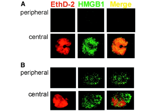
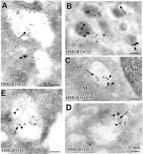
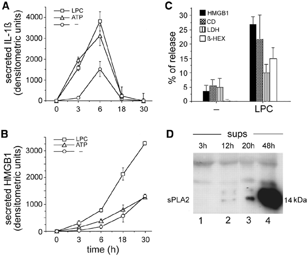
HMGB1, a non-histone nuclear factor, acts extracellularly as a mediator of delayed endotoxin lethality, which raises the question of how a nuclear protein can reach the extracellular space. We show that activation of monocytes results in the redistribution of HMGB1 from the nucleus to cytoplasmic organelles, which display ultrastructural features of endolysosomes. HMGB1 secretion is induced by stimuli triggering lysosome exocytosis. The early mediator of inflammation interleukin (IL)-1beta is also secreted by monocytes through a non-classical pathway involving exocytosis of secretory lysosomes. However, in keeping with their respective role of early and late inflammatory factors, IL-1beta and HMGB1 respond at different times to different stimuli: IL-1beta secretion is induced earlier by ATP, autocrinally released by monocytes soon after activation; HMGB1 secretion is triggered by lysophosphatidylcholine, generated later in the inflammation site. Thus, in monocytes, non-classical secretion can occur through vescicle compartments that are at least partially distinct.


Figures

Figure 1
Figure 1
Immunofluorescence analysis of HMGB1 in resting and LPS-activated monocytes. Monocytes, freshly isolated (A) or cultured for 18 h with LPS (B), were fixed, permeabilized, stained with ethidium homodimer-2 (EthD-2, red channel) and anti-HMGB1 antibody (HMGB1, green channel) and analyzed by confocal microscopy. Two sections of the same cells (peripheral and central) are shown, which allows the nuclear (central) and cytoplasmic (peripheral, absent at time 0, and evident after 18 h of activation) staining to be appreciated. The merged images (Merge) verify the almost complete colocalization of HMGB1 and EthD-2 at time 0 (A, yellow) which decreases after LPS activation (B).
Figure 2
Figure 2
Relocalization of HMGB1 in activated monocytes. (A) Aliquots of nuclei (lanes 1 and 2), postnuclear supernatants (PNS, lanes 3 and 4) and soluble cytosol (lanes 5 and 6) (corresponding to 5 × 10⁶ cells) from resting (t0, lanes 1, 3 and 5) or 18 h LPS-activated monocytes (t18, lanes 2, 4 and 6) were analyzed by western blotting with anti-HMGB1 antibody. One experiment out of six performed is shown. (B) Aliquots (50 μg) from P50 fractions from resting (t0, lanes 1 and 2) or activated monocytes (t18, lanes 3 and 4), treated with trypsin (Try, lanes 1 and 3) or with trypsin and Triton X-100 (TryTx, lanes 2 and 4) were analyzed by western blotting with anti-HMGB1 (upper), anti-IL-1β (middle) or anti-CD (lower) antibodies. One experiment of six performed is shown. (C) Confocal microscopy analysis of LPS-activated monocytes, double stained with anti-HMGB1 (red channel) and anti-EEA1 (green channel, left) or anti-LAMP1 (green channel, right) antibodies.
Figure 3
Figure 3
Immunoelectron microscopy analysis of freshly isolated (A) or 24 h activated monocytes (B and C), immunogold labeled for HMGB1. Arrows point to HMGB1 staining, restricted to the nucleus in (A) and localizing into vesicles in (B) and (C). In activated monocytes, HMGB1 appears associated to dense material in structures displaying the morphology of endosomes (C). The partial loss of the dense matrix is probably due to the fixation technique used to allow staining with anti-HMGB1 (see Methods), which led to partial extraction of vesicle content. Counting of gold particles allowed us to determine that, in resting monocytes (A), 65% of HMGB1 is in the nucleus and 35% in the cytoplasm; in activated monocytes (B and C), 26% of gold particles are nuclear and 74% are cytoplasmic, associated to endosomal structures.
Figure 4
Figure 4
Double immunogold labeling of HMGB1 and CD (A and B) and HMGB1 and IL-1β (CE). Arrows point to small gold particles (HMGB1), and arrowheads point to large gold particles (CD in A and B, IL-1β in C–E)
Figure 5
Figure 5
Kinetics of basal and induced secretion of mature IL-1β (17 kDa) and HMGB1. Densitometric analysis of IL-1β (A) and HMGB1 (B) secreted at different times in the absence of stimuli (circles) or after the addition of 1 mM ATP (triangles) or 20 μM LPC in the last 10 min (squares). The mean of a triplicate experiment ± SD is shown. One experiment of six perfomed is shown. (C) Release of HMGB1, CD, LDH and β-hexosoaminidase by 18 h LPS-activated monocytes, non-stimulated (−) or stimulated with 20 μM LPC for the last 10 min. Data are expressed as a percentage of secretion according to the following formula: secreted protein/intracellular + secreted protein × 100 ± SD. One experiment of four performed is shown. (D) Supernatants (sups) from monocytes at different times after LPS stimulation as indicated were analyzed for the presence of sPLA2 by western blotting.

References

    1. Andrei C., Dazzi C., Lotti L., Torrisi M.R., Chimini G. and Rubartelli A. (1999) The secretory route of the leaderless protein IL-1β involves exocytosis of endolysosome-related vesicles. Mol. Biol. Cell, 10, 1463–1475. - PMC - PubMed
    1. Andrews N.W. (2000) Regulated secretion of conventional lysosomes. Trends Cell Biol., 10, 316–321. - PubMed
    1. Bianchi M.E. and Beltrame M. (2000) Upwardly mobile proteins. Workshop: the role of HMG proteins in chromatin structure, gene expression and neoplasia. EMBO rep., 1, 109–114. - PMC - PubMed
    1. Blott E.J. and Griffiths G.M. (2002) Secretory lysosomes. Nat. Rev. Mol. Cell Biol., 3, 122–31. - PubMed
    1. Bodin P. and Burnstock G. (1998) Increased release of ATP from endothelial cells during acute inflammation. Inflamm. Res., 47, 351–354. - PubMed

Publication types

MeSH terms