

Continuous left hemidiaphragm sign revisited: a case of spontaneous pneumopericardium and literature review
- PMID: 12231618
- PMCID: PMC1767382
- DOI: 10.1136/heart.88.4.e5
Continuous left hemidiaphragm sign revisited: a case of spontaneous pneumopericardium and literature review
Abstract
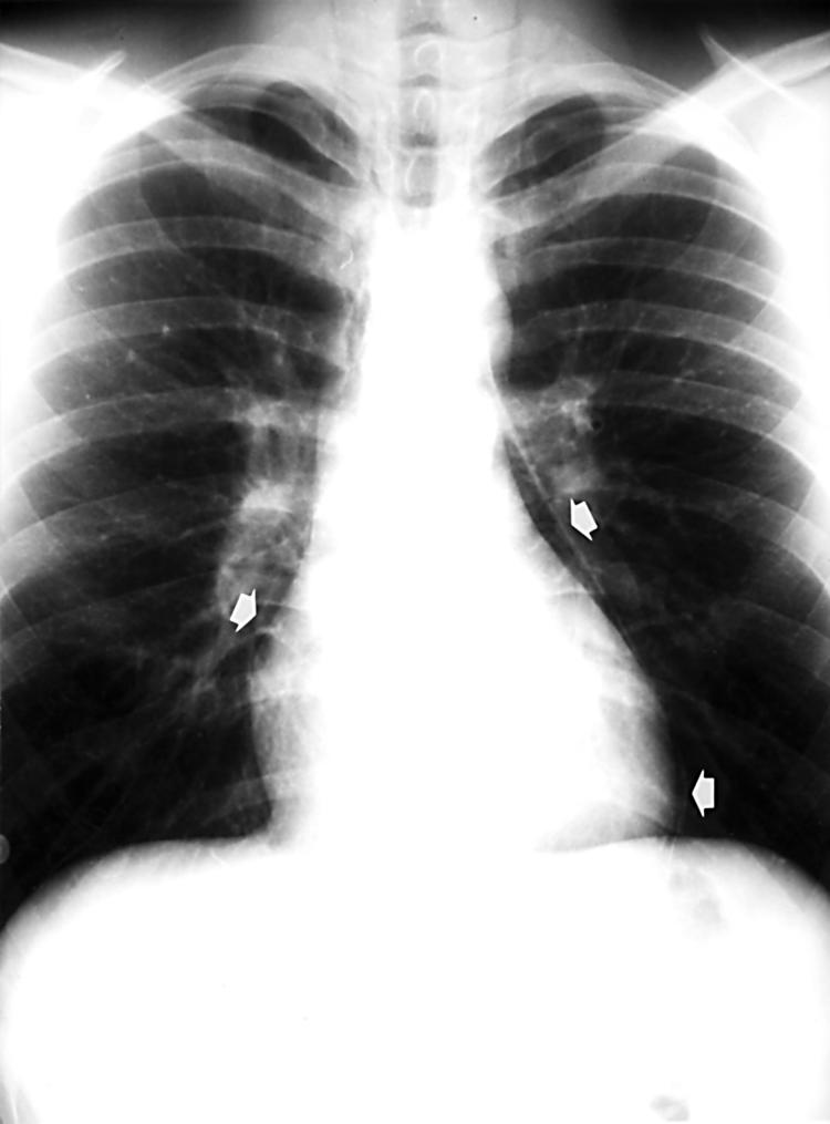
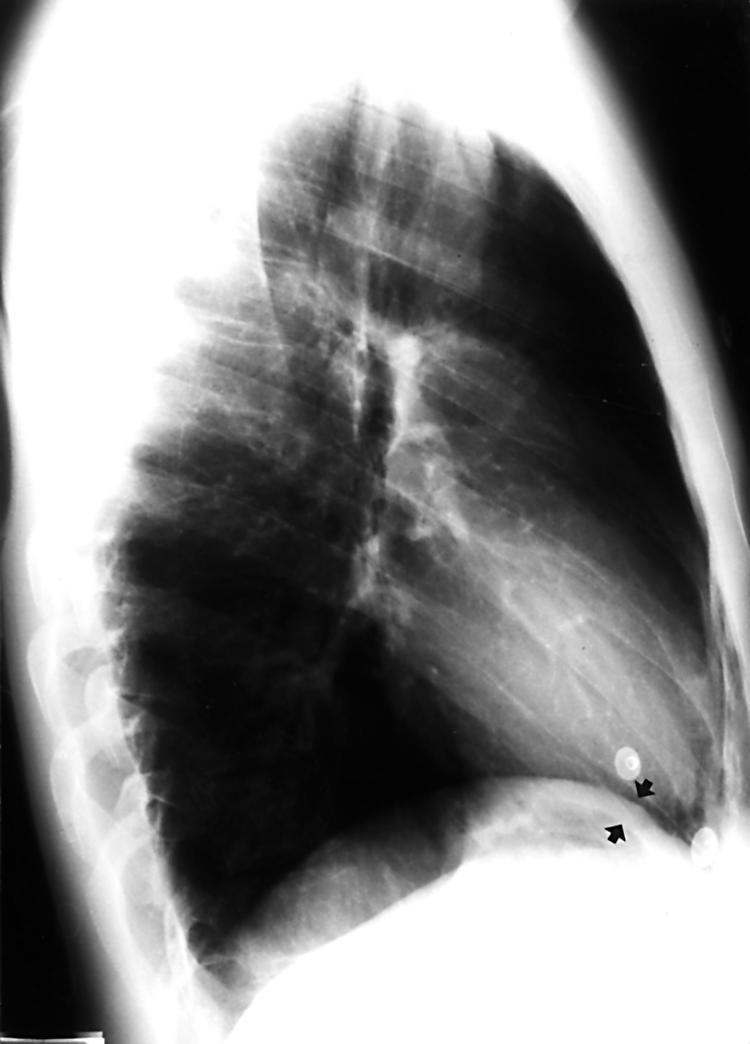
In pneumopericardium, a rare but potentially life threatening differential diagnosis of chest pain with a broad variety of causes, rapid diagnosis and adequate treatment are crucial. In upright posteroanterior chest radiography, the apical limit of a radiolucent rim, outlining both the left ventricle and the right atrium, lies at the level of the pulmonary artery and ascending aorta, reflecting the anatomical limits of the pericardium. The band of gas surrounding the heart may outline the normally invisible parts of the diaphragm, producing the continuous left hemidiaphragm sign in an upright lateral chest radiograph. If haemodynamic conditions are stable, the underlying condition should be treated and the patient should be monitored closely. Acute haemodynamic deterioration should prompt rapid further investigation and cardiac tamponade must be actively ruled out. Spontaneous pneumopericardium in a 20 year old man is presented, and its pathophysiology described.
Figures
References
-
- Bricketeau M. Observation d’hydropneumopéricarde accompagné d’un bruit de fluctuation perceptible a l’oreille. Arch Gen Med 1844;4:334.
-
- Tucker WS Jr. Symptoms and signs of syndromes associated with mill wheel murmurs. N C Med J 1988;49:569–72. - PubMed
-
- Capizzi PJ, Martin M, Bannon MP. Tension pneumopericardium following blunt injury. J Trauma 1995;39:775–80. - PubMed
-
- Reed JR, Thomas WP. Hemodynamics of progressive pneumopericardium in the dog. Am J Vet Res 1984;45:301–7. - PubMed
-
- Mauer ER, Mendez FL, Finkelstein M, et al. Cardiovascular dynamics in pneumopericardium and hydropericardium. Angiology 1958;9:176–9. - PubMed
Publication types
MeSH terms
LinkOut - more resources
Full Text Sources
Medical