Comparative Study
. 2002 Sep 15;16(18):2390-402.
doi: 10.1101/gad.1012602.

Regulation of sex-specific differentiation and mating behavior in C. elegans by a new member of the DM domain transcription factor family

Affiliations
Comparative Study

Regulation of sex-specific differentiation and mating behavior in C. elegans by a new member of the DM domain transcription factor family

Robyn Lints et al. Genes Dev. .

Abstract

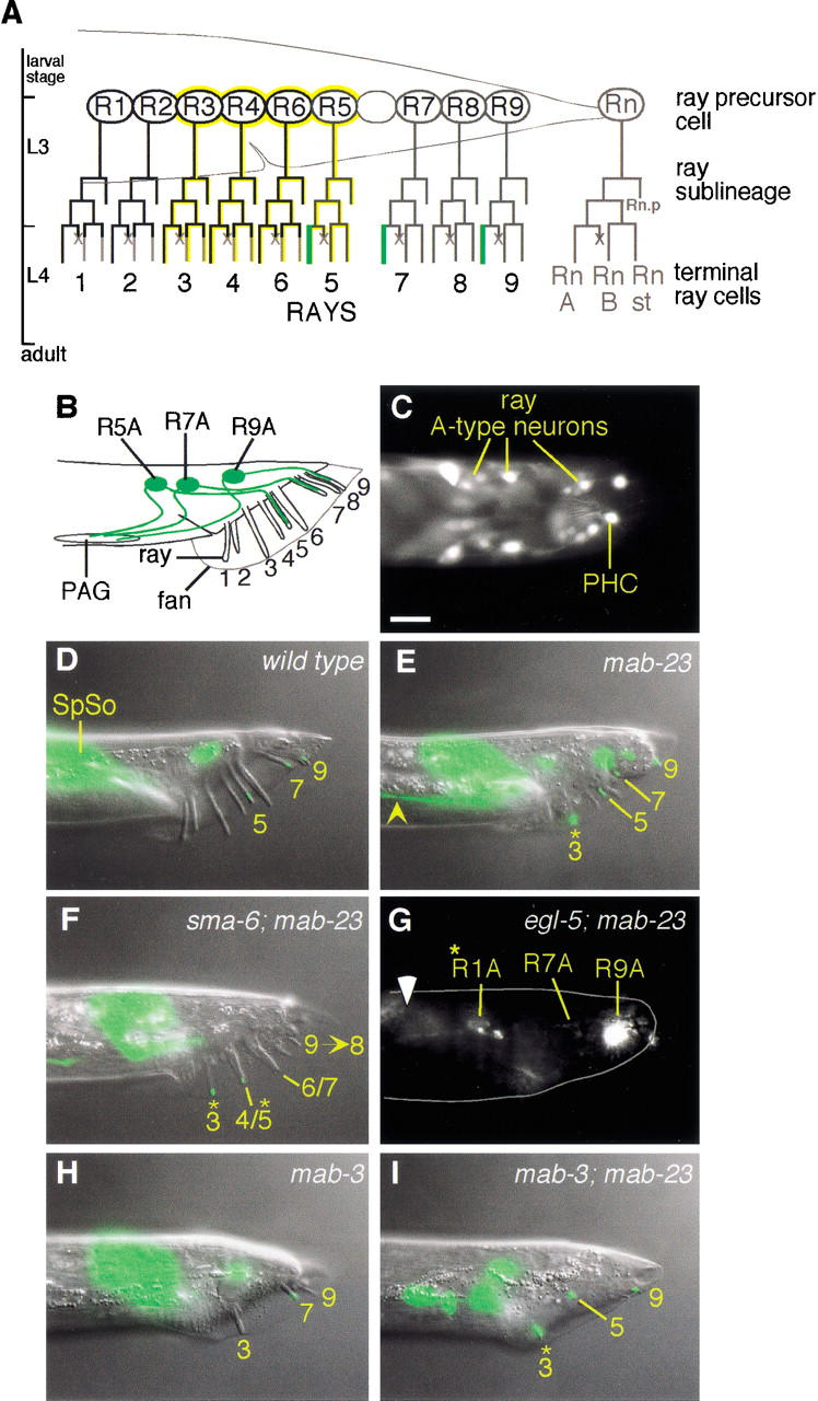
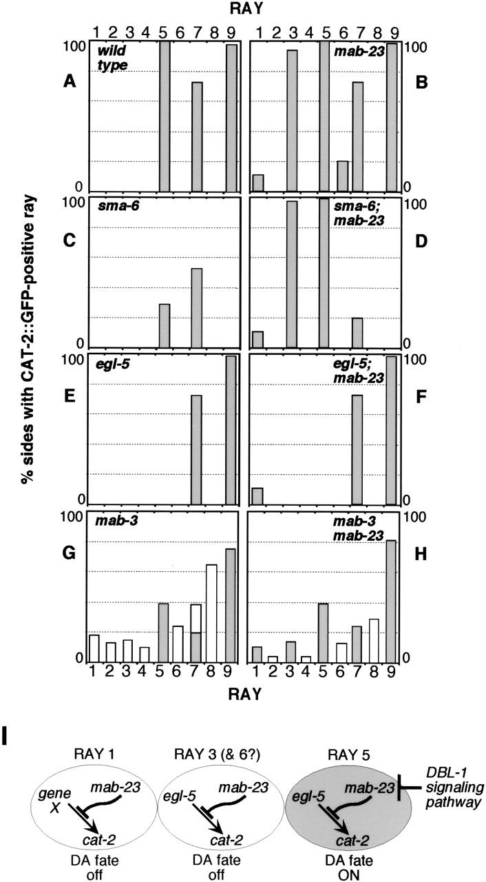
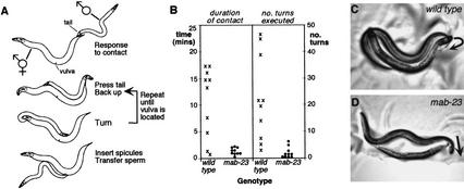
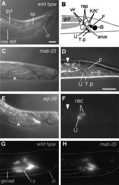
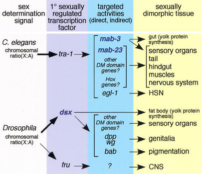
Mutations in Caenorhabditis elegans gene mab-23 cause abnormal male tail morphology and abolish male fecundity but have no obvious effect in the hermaphrodite. Here we show that mab-23 encodes a DM (Doublesex/MAB-3) domain transcription factor necessary for specific aspects of differentiation in sex-specific tissues of the male. mab-23 is required for the patterning of posterior sensory neurons in the male nervous system, sex muscle differentiation, and morphogenesis of the posterior hypodermis, spicules, and proctodeum. Failure of mab-23 mutant males to sire progeny is due primarily to defective sex muscle-mediated turning during copulatory behavior and likely compounded by impairment of sperm passage through the proctodeum. In the male nervous system, mab-23 refines ray neuron subtype distribution by restricting expression of dopaminergic neurotransmitter identity through interactions with the Hox gene egl-5 and a TGF-beta-related signaling pathway. mab-23 has distinct roles and functions independent of mab-3, indicating different aspects of C. elegans male sexual differentiation are coordinated among DM domain family members. Our results support the hypothesis that DM domain genes derive from an ancestral male sexual regulator and suggest how regulation of sexual development has evolved in distinct ways in different phyla.


Figures

Figure 1
Figure 1
mab-23 encodes a DM domain protein. (A) Genetic map of a portion of linkage group V (top) and the corresponding physical map showing the minimal 9.5-kb mab-23 rescuing region (middle), common to cosmids C32C4 and H09F14, and the predicted mab-23 ORF, g-V-2130 (bottom). The positions of mab-23 mutations and site of the GFP ORF insertion in mab-23 reporter EM#308 is indicated. (B) Sequence alignment of DM domains. Cysteines and histidines that form the Zn2+-binding sites are indicated (boxed red or blue; for review, see Zhu et al. 2000). Percentage identity with MAB-23 is shown; identical amino acids are indicated in black; similar amino acids in grey. MAB-23 (GenBank accession no. AF535153); MAB-3a, and MAB-3b, the two DM domains of C. elegans MAB-3 (accession no. O18214); Drosophila DSX (accession no. P23022/P23023); human DMRT1 (accession no. AF130728); zebrafish TERRA (accession no. AF080622). e2518 converts the last conserved cysteine of the DM motif to phenylalanine. bx118 alters the fifth position of the 5′ donor splice site within intron 1. (C) Unrooted phylogenetic tree of DM domains encoded by predicted genes in human (hu, black), Drosophila melanogaster (Dm, blue), and C. elegans (Ce, red) genomes. The DM domains (zinc module plus recognition helix) were aligned and subjected to phylogenetic analysis by maximum parsimony using the Protpars algorithm of PHYLIP v. 3.573c (Felsenstein 1989). The tree presented is a consensus of the most parsimonious trees derived from 100 bootstrap resamplings of the original data. Bootstrap support values over 50% are shown; nodes with values of <50% were collapsed. DM domains of known sexual regulator genes are boxed. Accession numbers are as follows: human DMRTA1 (AJ290954), DMRTA3 (AAF78891), DMRTA2 (AJ301580), DMRTC2 (AJ291669), DMRTB1 (AJ291671), DMRT2 (AF130729); Drosophila dmrt93B (AAF55843), dmrt11E (AAF48261), dmrt99B (AAF56919); C. elegans C27C12.6 (CAA93739), C34D1.1 (CAB01491), C34D1.2 (CAB01490), F10C1.5 (AAA93409), F13G11.1 (CAB05898/CAB05899), K08B12.2 (AAB52258), T22H9.4a and T22H9.4b, the two DM domains of T22H9.4 (AAC69225), Y43F8C.10 (CAA21612), and Y67D8A.3 (AAK68545).
Figure 2
Figure 2
mab-23 is required for ray dopaminergic patterning and axon pathfinding. (A) Postembryonic cell lineages leading to the ray neurons (Sulston and Horvitz 1977). RnA, A-type neuron; RnB, B-type neuron; Rnst, structural cell; ×, programmed cell death. Rn.p is a hypodermal cell. Dopaminergic A-type neurons (green; Sulston and Horvitz 1977), EGL-5-expressing cells (yellow; Ferreira et al. 1999), MAB-23 ∷ GFP-expressing cells (black). (B) Adult male tail (lateral view; anterior, left; dorsal, up) showing the location of dopaminergic ray neurons (green). PAG, preanal ganglion. (C) mab-23 expression in wild-type adult male rays. Fluorescent micrograph of a transgenic adult male (dorsal view) expressing EM#308 reporter. MAB-23 ∷ GFP localizes predominantly to cell nuclei in the A-type neurons of rays 1–4 and 6 and the nonsex-specific PHCL/R neurons. (DI) Dopaminergic fate expression in wild-type and mutant males (lateral view; anterior, left; dorsal, up). The dopaminergic fate marker is CAT-2 ∷ GFP (Lints and Emmons 1999). Nomarski and fluorescent micrographs of the same animal are superimposed. (D) Wild type. CAT-2 ∷ GFP is visible in ray 5, 7, and 9 A-type neurons. CAT-2 ∷ GFP signal more anteriorly in the tail corresponds to male spicule socket cell (SpSo) expression. (E) mab-23(bx118). The ray 3 A-type neuron expresses CAT-2 ∷ GFP ectopically (*) and its axon projects abnormally (yellow arrowhead). The Morpho-Mab tail defect characteristic of mab-23 males is also apparent in this animal. (F) sma-6(wk7); mab-23(bx118). Ray abnormalities typical of DBL-1 pathway single mutants are apparent: fusion of rays 4 and 5 (4/5) and rays 6 and 7 (6/7), and ray 9 has been transformed to ray 8 (9→8). CAT-2 ∷ GFP is ectopically expressed in rays 3 and 4/5. (G) egl-5(u202); mab-23(bx118). Fluorescent micrograph [the presence of egl-5(u202) disrupts male tail retraction and, therefore, rays and fan are embedded in the tail]; body outline (gray solid line); gut autofluorescence (white arrowhead). CAT-2 ∷ GFP is absent form the A-type neurons of egl-5-dependent rays 3 and 5. CAT-2 ∷ GFP is present in the A-type neurons of non-egl-5-dependent rays 7 and 9 (R7A and R9A, respectively) and ectopically in ray 1 (R1A*). (H) mab-3 (e1240). Only rays 3, 7, and 9 are generated in this animal. CAT-2 ∷ GFP expression is observed in ray 7, one of the rays that express this fate in wild type. No ectopic expression of CAT-2 ∷ GFP is observed in the ray 3 that is present. (I) mab-3 (e1240); mab-23 (bx118). Double mutant shows both mab-3 and mab-23 phenotypes: missing rays and ectopic expression of CAT-2 ∷ GFP in ray 3, respectively. Magnification, 1000×. Bar, 10 μm.
Figure 3
Figure 3
Genetic interactions between mab-23, egl-5, and the DBL-1 pathway. (A–H) Frequency and distribution of CAT-2 ∷ GFP-positive rays in wild-type and mutant males. Single, double, and triple mutant strains were generated by crossing either wild-type, mab-23(bx118), or mab-23(e2518);nIs118 animals with DBL-1 pathway, egl-5, or mab-3 mutants. The DBL-1 pathway is defined by the six genes, dbl-1, daf-4, sma-6, sma-2, sma-3, and sma-4 (Patterson and Padgett 2000); for mutations used in this study, see Materials and Methods. sma-6(wk7), egl-5(u202), and mab-3(e1240) are presumptive null mutations (Wang et al. 1993; Raymond et al. 1998; Krishna et al. 1999). CAT-2 ∷ GFP-expressing rays were identified as described in Lints and Emmons (1999). mab-23(bx118) and mab-23(e2518) mutant combinations gave similar results, as did mutations in all genes of the DBL-1 pathway. Tail sides develop independently and are, therefore, scored separately. The number of sides scored in a given mutant background ranged from 60 to 144. For each genetic background, the pattern of CAT-2 ∷ GFP observed was found to be consistent with the dopamine staining pattern obtained using FIF (Sawin et al. 2000; data not shown). In DBL-1 pathway mutant backgrounds, fusions involving rays 5, 7, or 9 have been grouped with unfused ray 5, 7, or 9 identities, respectively, as fusions contain a single CAT-2 ∷ GFP-positive neuron likely to correspond to the neuron that normally expresses CAT-2 ∷ GFP in wild type, namely the A-type neuron of ray 5, 7, or 9 (Lints and Emmons 1999). In mab-3 single and mab-3; mab-23 double mutants, rays 1–6 are variably missing so that the frequency with which a particular ray identity was generated is indicated (unshaded), in addition to the frequency with which it was CAT-2 ∷ GFP-positive (shaded). (I) Genetic relationship between mab-23, the DBL-1 pathway, and egl-5 in dopaminergic (DA) patterning (see text).
Figure 4
Figure 4
mab-23 males are defective in specific steps of male mating behavior. (A) Wild-type C. elegans male mating behavior. The male responds to contact with the hermaphrodite by halting forward locomotion, placing the ventral side of his tail against the hermaphrodite's body and swimming backward in search of the vulva. If the male reaches the end of her body without finding the vulva he executes a turn to continue the search on the other side. After locating the vulva, he inserts anchoring spicules and transfers sperm. (B) Performance of wild-type () and mab-23 (•) males in mating assays. Ten adult virgin males of each genotype were individually observed and scored for the amount of time spent in direct contact with the hermaphrodite (left) and for the number of successful turns executed (right) during the first 30 min of a mating assay (see Materials and Methods). (C,D) Examples of posture adopted by wild-type (C) and mab-23 (D) males during execution of the turn. Trajectory of the turn is indicated by the direction of the arrow. The posture adopted by wild-type males ensures that the ventral side of the male tail is pressed against the hermaphrodite throughout the turn. mab-23 males do not adopt this posture and execute loose or missed turns (shown here), during which contact with the hermaphrodite is lost.
Figure 5
Figure 5
mab-23 is required for male-specific muscle cell differentiation. (A,B) MAB-23 ∷ GFP is expressed in ventral posterior muscles of the male. Fluorescent micrographs of MAB-23 ∷ GFP in non-sex-specific ventral body wall (b.w.) muscle, male-specific outer longitudinal (o.l.), and diagonal muscles (lateral view, A) and diagonal muscles (ventral view, B). (C,D) Affect of exogenous 20 mM 5HT on wild-type and mab-23 males (lateral view). (C) Wild type. Note tight ventral flexure of the tail (white arrowhead) and altered posture affecting more anterior regions of the body. (D) mab-23. Ventral flexure of the tail is weak and more anterior regions of the body also differ in posture from wild type. (E,F) SER-2 ∷ GFP expression in the diagonal muscles. Fluorescent micrographs showing strong expression of SER-2 ∷ GFP in the diagonal muscles (arrowheads) of a wild-type male (E; ventral view) and weak expression in a mab-23 male (F; ventral view). v.n.c., ventral nerve cord. (G) Frequency of 5HT-induced male tail curling. The percentage of males that displayed a tight ventral flexure of the tail (as shown in C) after exposure to 20 mM 5HT is shown. Number of males scored were as follows: wild-type, 70; mab-23(bx118), 43; mab-23(e2518), 50; and mab-3(e1240), 40. (H) Mean number of diagonal muscles per male side that were SER-2 ∷ GFP-positive and have wild-type morphology (revealed by UNC-27 ∷ GFP labeling) for the genotype indicated is shown, as well as the range observed in that population. In both mab-23 and mab-3 males, SER-2 ∷ GFP signal is also weak (as shown in F) compared with wild-type. Number of male sides scored (SER-2 ∷ GFP, morphology) were as follows: wild-type (49, 30); mab-23(bx118) (48, 39); mab-23(e2518) (48, 43); and mab-3(e1240) (30, 49). Magnification: A, 400×; B,E,F, 1000×. Bar, 10 μM.
Figure 6
Figure 6
mab-23 is required for development of the male proctodeum. (A,C,E) Proctodeum of wild-type, mab-23 and egl-38 adult males. Nomarski micrographs (lateral view; anterior, left; dorsal, up). (A) In wild-type males the vas deferens (vas def.) is devoid of sperm. cl., cloaca (the anus in larval animals); sp., spicules. In mab-23 (C) and egl-38 (E) males, sperm (arrowhead) accumulates in the vas def. (B) Schematic of L1 male hindgut (lateral view). Black, blast cells; vir, rectal valve cells; rep, rectal epithelial cells (Sulston et al. 1980). (D) Nomarski micrograph of L1 male hindgut (lateral view; anterior, left; dorsal, up). (F) Fluorescent micrograph of D showing MAB-23 ∷ GFP in U and two rectal epithelial cells (rec), either K, K.a, K‘, or repD; gut autofluorescence (white arrowhead). (G,H) Fluorescent micrograph showing plin-48 ∷ gfp-expressing hindgut cells in late L4 males (lateral view). U descendents and K‘/K.a are located over the posterior end of the gonad (outlined with broken line) in the ventral half of the animal. In wild-type (G), these cells extend anteriorly over the gonad; in mab-23 males (H), these cells show only limited extension. Body outline (solid line), engulfed linker cell corpse inside U.l/rp (l.c). Magnification, 1000×. Bar, 10 μM.
Figure 7
Figure 7
Hierarchy of sexual differentiation genes in C. elegans and Drosophila. In both C. elegans and Drosophila, the primary sex determining signal, the X chromosome to autosome ratio (X : A), sets the activity of the globally acting sex determination pathway. DM domain genes (blue) in C. elegans and Drosophila differ with respect to their relationship to this pathway. In C. elegans, the primary sexually regulated transcription factor is a Gli-related Zn finger protein, TRA-1A, encoded by the terminal gene of the sex determination pathway, tra-1. tra-1 activity affects the development of most sexually dimorphic tissues, acting through non-sex-specific developmental gene targets (e.g., cell death gene egl-1), as well as dedicated sexual regulators (mab-3). mab-3 (in tissues other than the gut) and mab-23 are not directly targeted by TRA-1A. The sex specificity of their activity derives from the cellular context in which they are expressed. In Drosophila, the primary sexually regulated transcription factor is the DM domain gene dsx. In contrast to C. elegans DM domain genes, dsx is a direct target of the sex-determination pathway in most somatic tissues. Like tra-1, dsx is broadly acting and functions early in sexual differentiation to sex-specifically regulate multiple developmental genes. In the CNS, fru appears to be the primary sexually regulated transcription factor.

Comment in

Similar articles

Cited by

References

    1. Altun-Gultekin Z, Andachi Y, Tsalik EL, Pilgrim D, Kohara Y, Hobert O. A regulatory cascade of three homeobox genes, ceh-10, ttx-3 and ceh-23, control cell fate specification of a defined interneuron class in C. elegans. Development. 2001;128:1951–1969. - PubMed
    1. Baker BS, Ridge K. Sex and the single cell. I. On the action of major loci affecting sex determination in Drosophila melanogaster. Genetics. 1980;94:383–423. - PMC - PubMed
    1. Barr MM, Sternberg PW. A polycystic kidney-disease gene homologue required for male mating behaviour in C. elegans. Nature. 1999;401:386–389. - PubMed
    1. Belote JM, Handler AM, Wolfner MF, Livak KJ, Baker BS. Sex-specific regulation of yolk protein gene expression in Drosophila. Cell. 1985;40:339–348. - PubMed
    1. Brenner S. The genetics of Caenorhabditis elegans. Genetics. 1974;77:71–94. - PMC - PubMed

Publication types

MeSH terms

Substances

LinkOut - more resources