. 2002 Sep 15;16(18):2415-27.
doi: 10.1101/gad.1009002.

Combinatorial control of Drosophila eye development by eyeless, homothorax, and teashirt

Affiliations

Combinatorial control of Drosophila eye development by eyeless, homothorax, and teashirt

Jose Bessa et al. Genes Dev. .

Abstract

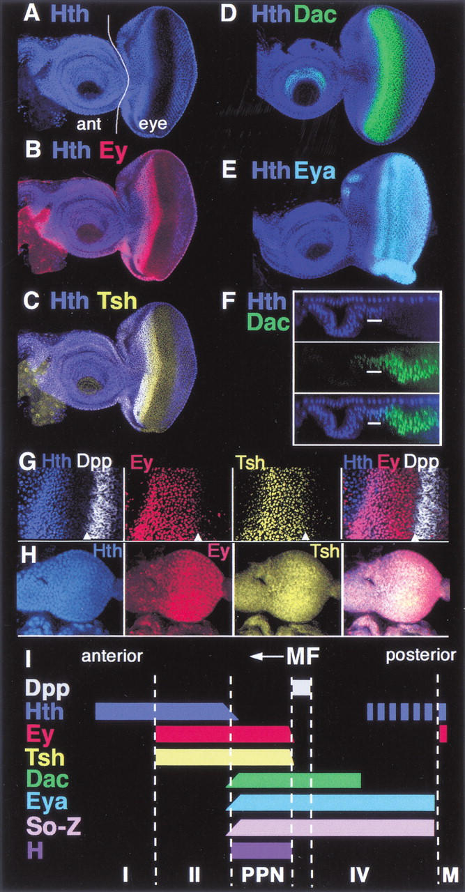
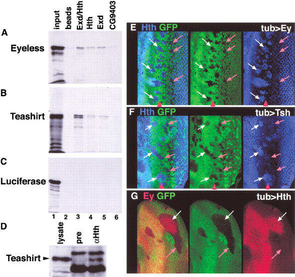
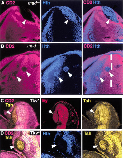
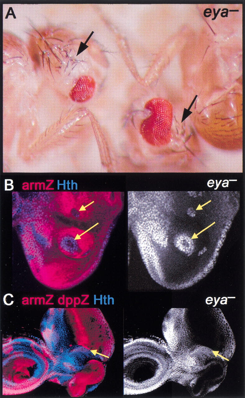
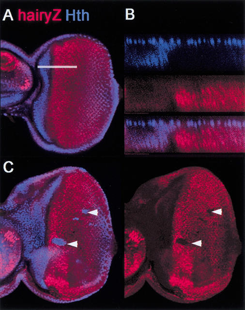
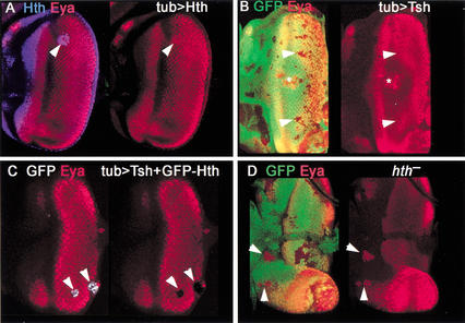
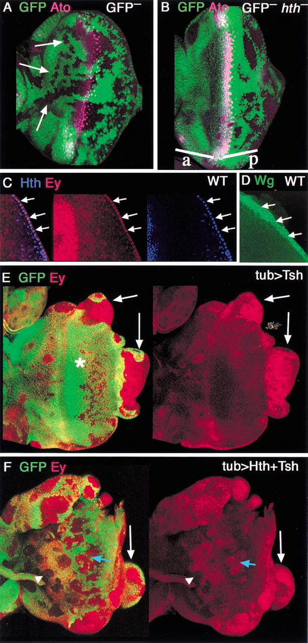
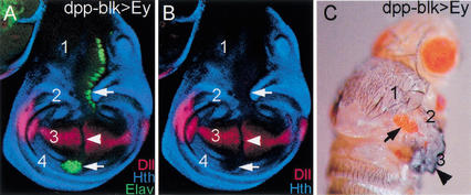
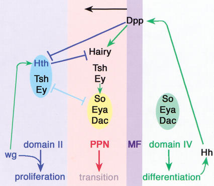
In Drosophila, the development of the compound eye depends on the movement of a morphogenetic furrow (MF) from the posterior (P) to the anterior (A) of the eye imaginal disc. We define several subdomains along the A-P axis of the eye disc that express distinct combinations of transcription factors. One subdomain, anterior to the MF, expresses two homeobox genes, eyeless (ey) and homothorax (hth), and the zinc-finger gene teashirt (tsh). We provide evidence that this combination of transcription factors may function as a complex and that it plays at least two roles in eye development: it blocks the expression of later-acting transcription factors in the eye development cascade, and it promotes cell proliferation. A key step in the transition from an immature proliferative state to a committed state in eye development is the repression of hth by the BMP-4 homolog Decapentaplegic (Dpp).


Figures

Figure 1
Figure 1
Transcription factor expression domains in the eye imaginal disc. All images are of wild-type eye–antennal discs. Anterior is to the left. (AE) Eye–antennal discs stained for (A) Hth, (B) Hth + Ey, (C) Hth + Tsh, (D) Hth + Dac, and (E) Hth + Eya as indicated. In A the antennal (ant) and eye portions of the disc are indicated. (F) An optical cross-section of a disc stained for Hth and Dac, showing the reciprocal relationship between these two factors in the main epithelium of the eye disc. Eya and Hth have a similar relationship (data not shown). The upper row of Hth-positive nuclei are in the peripodial membrane. (G) Closeup of the anterior region of a disc stained for Hth, Ey, Tsh, and dpp–lacZ. The arrowheads point to the anterior edge of the MF, as marked by dpp–lacZ expression. Although Tsh and Ey are not expressed posterior to the furrow, some background signal is observed in these images. (H) Second-instar eye–antennal discs stained for Hth, Ey, and Tsh. Ey is expressed evenly throughout the eye disc. Hth and Tsh are also expressed in all cells, but at lower levels posteriorly. (I) A summary of these expression patterns. The expression domains of Hairy (H) and Dpp are also summarized. The So-Z pattern is based on a lacZ enhancer trap into so (data not shown). Based on these patterns, the following domains can be defined: domain I (Hth, alone); domain II (Hth, Tsh, Ey); PPN (Tsh, Ey, So, Eya, Dac, H); domain IV (So, Eya, Dac); MF (morphogenetic furrow); and M (margin cells, Hth, Ey).
Figure 2
Figure 2
In vitro and in vivo interactions among Ey, Hth, and Tsh. (AC) In vitro interaction experiments. (A) 35S-Ey, (B) 35S-Tsh, or (C) 35S-Luciferase proteins pulled down with empty beads (lane 2), an Hth/Exd dimer (lane 3), Hth alone (lane 4), Exd alone (lane 5), or CG9403, a zinc-finger protein that serves as a negative control (lane 6). The input for each 35S protein is shown in lane 1. (D) Total embryo extracts were immunoprecipitated with either preimmune sera (pre) or anti-Hth sera (αHth), run on an SDS-PAGE gel, and probed with an anti-Tsh antibody. A total embryonic lysate shows the position of Teashirt (lysate). (EG) The MF region of eye discs containing clones of cells expressing Ey (E), Tsh (F), or Hth (G). Anterior is to the left, and the red arrowheads point to the approximate position of the MF. Clones are marked by the absence of GFP (green) and were stained for Hth (E,F) or Ey (G). White arrows point to regions that show ectopic expression and pink arrows point to regions that do not show ectopic expression.
Figure 3
Figure 3
hth, but not ey or tsh, is repressed by Dpp in the eye disc. (A,B) mad clones, marked by the absence of CD2 (fucsia), stained for Hth (blue). Arrowheads point to clones. The dashed line in B marks the approximate position of the MF. (C,D) Clones expressing Tkv*, marked by the absence of CD2 (fucsia) and stained for (C) Tsh + Ey or (D) Tsh + Hth.
Figure 4
Figure 4
eya represses hth and head capsule development. (A) Adult flies containing eya clones. Although eya tissue is unmarked, these heads show a loss of eye and a corresponding increase in head capsule (arrows). (B,C) eya clones, marked by the absence of arm–lacZ (red), stained for Hth (blue in left panels and white in right panels). Hth is de-repressed cell-autonomously in eya cells. In C, the disc also expresses dpp–lacZ (strong red stripe). Hth is de-repressed in eya cells, even when they are adjacent to Dpp-expressing cells (yellow arrows in C).
Figure 5
Figure 5
Hth represses hairy. (A) Wild-type eye disc stained for Hth (blue) and Hairy–lacZ (red). Note that Hairy–lacZ is observed more posteriorly than Hairy because of persistence of β-galatosidase (data not shown). (B) An optical cross-section of a wild-type disc (at the position of the white line in A), showing no overlap between Hth and Hairy. (C) Ectopic clones of Hth (arrowheads), stained for Hth (blue) and Hairy–lacZ. Hth represses Hairy–lacZ expression.
Figure 6
Figure 6
Ey + Hth + Tsh represses eya. (A) Clone of ectopic GFP–Hth in an eye disc (arrowhead) stained for Hth (blue) and Eya (red). No repression of Eya is observed. (B) Clones of ectopic Tsh, marked by the absence of GFP, in an eye disc stained for Eya (red). No repression is observed (arrowheads). The * marks an overgrowing clone in the peripodial membrane in a different focal plane that causes a distortion of the eye disc epithelium. (C) Clones of ectopic Tsh + GFP–Hth stained for GFP (white) and Eya (red). Repression of Eya is observed (arrowheads). (D) hth clones, marked by the absence of GFP, stained for Eya. De-repression of Eya is observed (arrowheads). De-repression of dac is also observed in hth clones (data not shown).
Figure 7
Figure 7
Genetic control of proliferation of the eye disc. (A,B) Neutral (A) or hth (B) clones, marked by the absence of GFP, and stained for the MF marker, Ato (fuchsia). Neutral clones are observed both anterior (arrows) and posterior to the MF. hth clones are only rarely observed anterior (a) to the MF but are readily observed posterior (p) to the furrow. (C) Detail of the posterior margin of a wild-type eye disc stained for Hth (blue) and Ey (red). Hth and Ey are both expressed in these cells (arrows). Tsh is not expressed in these cells (data not shown). (D) Similar region of a wild-type disc stained for Wg (green), which is expressed in margin cells. (E,F) Clones of cells expressing (E) Tsh or (F) Tsh + Hth, marked by the absence of GFP, stained for Ey (red). (E) Tsh clones grow poorly in the middle of the disc (*) but can induce large overgrowths at the edge of the disc (arrows). Overgrowing clones, but not internal clones, express Ey. (F) Tsh + Hth clones grow well in the middle of the disc (blue arrow) and also induce overgrowths at the edge of the disc (white arrow). Most clones maintain Ey expression, although some anterior clones show repression of Ey (arrowhead).
Figure 8
Figure 8
Induction of ectopic eyes in the hinge region of wing discs. (A,B) Wing imaginal disc in which Ey was expressed using (A) dppblk–Gal4, stained for Hth (blue), Elav (green), and Distalless (Dll; red); (B) only the Hth and Dll channels are shown. dppblk–Gal4 is expressed in a dorsal-to-ventral stripe across the entire wing disc just anterior to the AP compartment boundary. Wing discs have four main regions: (1) notum, (2) dorsal hinge, (3) wing pouch, and (4) ventral hinge. Hth is expressed in regions 1, 2, and 4, but not in 3. Tsh is expressed in regions 1, 2, and 4 also. Ectopic eye tissue, as detected by Elav, is observed in regions 1, 2, and 4 (arrows), but not in 3 (arrowhead). Dll and Hth are repressed by Ey expression in regions 1, 2, 3, and 4. dppblk–Gal4; UAS–ey also generates ectopic eyes in the proximal leg disc (data not shown). A similar correlation between ectopic Elav and Hth + Tsh expression was observed with several other Gal4 lines driving Ey (data not shown). (C) Adult fly expressing Ey via the dppblk–Gal4 driver line, showing a transformation of the hinge to eye (arrow). Wing blade development is partially suppressed in these flies, although wing tissue is still formed (arrowhead).
Figure 9
Figure 9
Summary. The factors and their interactions present in domains II, IV, PPN, and the MF are schematized. Do main IV expresses the differentiation transcription factors So, Eya, and Dac. Differentiated cells also express Hh, which induces Dpp expression in the MF. Dpp represses Hth and induces Hairy. Hth represses Hairy. The combination of Hth–Tsh–Ey is mutually antagonistic with So–Eya–Dac. Ey also directly activates So expression (Niimi et al. 1999), perhaps in the PPN domain. Also indicated is a positive requirement for Wg signaling for Hth expression in domain II (data not shown). Wg, together with the factors present in domain II, is proposed to promote proliferation in the eye disc. Not indicated here is our observation that Eya and Dac collaborate with Dpp to repress Hth and our inference that an unknown short-range signal in the MF represses Tsh and Ey.

Similar articles

Cited by

References

    1. Abu-Shaar M, Mann RS. Generation of multiple antagonistic domains along the proximodistal axis during Drosophila leg development. Development. 1998;125:3821–3830. - PubMed
    1. Allen TD, Zhu YX, Hawley TS, Hawley RG. TALE homeoproteins as HOX11-interacting partners in T-cell leukemia. Leuk Lymphoma. 2000;39:241–256. - PubMed
    1. Azpiazu N, Morata G. Function and regulation of homothorax in the wing imaginal disc of Drosophila. Development. 2000;127:2685–2693. - PubMed
    1. Baker NE. Transcription of the segment-polarity gene wingless in the imaginal discs of Drosophila, and the phenotype of a pupal-lethal wg mutation. Development. 1988;102:489–497. - PubMed
    1. ————— Cell proliferation, survival, and death in the Drosophila eye. Semin Cell Dev Biol. 2001;12:499–507. - PubMed

Publication types

MeSH terms