. 2002 Sep 15;16(18):2428-42.
doi: 10.1101/gad.1011602.

Synapsis-dependent and -independent mechanisms stabilize homolog pairing during meiotic prophase in C. elegans

Affiliations

Synapsis-dependent and -independent mechanisms stabilize homolog pairing during meiotic prophase in C. elegans

Amy J MacQueen et al. Genes Dev. .

Abstract

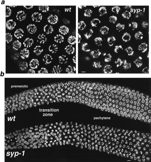
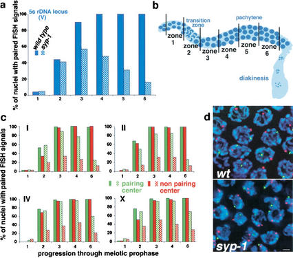
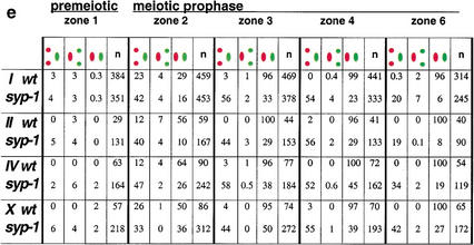
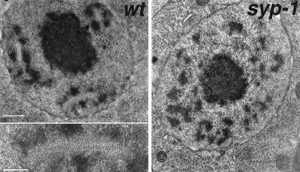
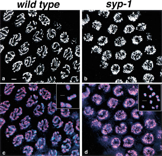
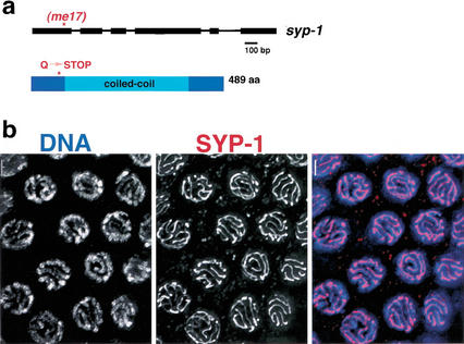
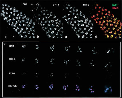
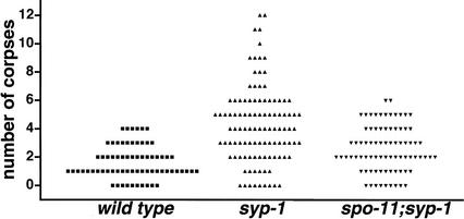
Analysis of Caenorhabditis elegans syp-1 mutants reveals that both synapsis-dependent and -independent mechanisms contribute to stable, productive alignment of homologous chromosomes during meiotic prophase. Early prophase nuclei undergo normal reorganization in syp-1 mutants, and chromosomes initially pair. However, the polarized nuclear organization characteristic of early prophase persists for a prolonged period, and homologs dissociate prematurely; furthermore, the synaptonemal complex (SC) is absent. The predicted structure of SYP-1, its localization at the interface between intimately paired, lengthwise-aligned pachytene homologs, and its kinetics of localization with chromosomes indicate that SYP-1 is an SC structural component. A severe reduction in crossing over together with evidence for accumulated recombination intermediates in syp-1 mutants indicate that initial pairing is not sufficient for completion of exchange and implicates the SC in promoting crossover recombination. Persistence of polarized nuclear organization in syp-1 mutants suggests that SC polymerization may provide a motive force or signal that drives redispersal of chromosomes. Whereas our analysis suggests that the SC is required to stabilize pairing along the entire lengths of chromosomes, striking differences in peak pairing levels for opposite ends of chromosomes in syp-1 mutants reveal the existence of an additional mechanism that can promote local stabilization of pairing, independent of synapsis.


Figures

Figure 1
Figure 1
Defective chromosome organization in syp-1 meiotic prophase nuclei. (a) High-magnification images of DAPI-stained nuclei from the pachytene region of wild-type and syp-1 mutant germ lines. Widely distributed about the periphery of wild-type pachytene nuclei are parallel pairs of DAPI-stained tracks, corresponding to lengthwise-aligned and synapsed homologous chromosomes. In the syp-1 mutant, pachytene region nuclei exhibit disorganized DAPI-stained tracks not aligned in parallel pairs, and chromatin is distributed asymmetrically within the nuclei. Images are projections approximately halfway through 3D data stacks of whole nuclei. (b) Low-magnification images of DAPI-stained germ lines. In both wild-type and syp-1 mutant germ lines, DAPI-stained chromatin is widely dispersed in premeiotic nuclei, whereas the polarized organization of nuclei in the transition zone (entering meiotic prophase) imparts a crescent-shaped appearance to DAPI-stained chromatin. In the pachytene region of the wild-type germ line, DAPI signals are again widely dispersed; early prophase nuclear polarization persists in the pachytene region of the syp-1 mutant germ line, with most nuclei in this region exhibiting an asymmetric distribution of chromatin. Images are projections of 3D data stacks that encompass entire nuclei (except at the edges, where only a portion of some nuclei are projected). Bars, a, 2 μm; b, 4 μm.
Figure 2
Figure 2
Homolog pairing is established but not maintained in syp-1 mutants. (a) Graph exhibiting the percent of nuclei with paired FISH signals at the 5S rDNA locus on Chromosome V. The numbers of nuclei scored per zone for wild-type and syp-1 (respectively) are as follows: zone 1: 103, 191; zone 2: 133, 242; zone 3: 125, 235; zone 4: 120, 219; zone 5: 102, 286; zone 6: 84, 148. In a and c, solid bars correspond to wild-type pairing frequencies, whereas hatched bars represent pairing frequencies in the syp-1 mutant. (b) Diagram of the germ line, indicating the positions of zones used in time-course analysis of homolog pairing. Zone 1, which begins approximately three nuclear diameters from the distal tip, contains exclusively premeiotic nuclei; zone 2 includes some premeiotic nuclei, but contains predominantly nuclei at the leptotene/zygotene stages of meiotic prophase; zones 3 and 4 contain mainly early- to mid-pachytene stages (zone 3 may also include a few leptotene/zygotene nuclei); zones 5 and 6 contain late-pachytene nuclei (zone 5 was used only for the experiment shown in a). (c) Graphs exhibiting data from double-label experiments in which pairing at the pairing-center end (green) and the non-pairing-center end (red) of a single chromosome were quantified simultaneously. For Chromosome I, the non-pairing-center end locus is ∼15% of the physical chromosome length from the left end of I; in all other cases, probes targeted loci <3% of chromosome length from the nearest chromosome end. Numbers of nuclei scored for each zone are exhibited in e; see Materials and Methods for statistical analysis. (d) High-magnification images of zone 4 nuclei hybridized with FISH probes targeting opposite ends of Chromosome IV (scale bar, 2 μm). FISH signals for both probes are paired in all wild-type nuclei. In the syp-1 mutant, the non-pairing-center end (red) is frequently unpaired, whereas the pairing-center end (green) is usually paired. (e) Percent of nuclei with distinct pairing configurations (e.g., green paired, red unpaired) within zones 1–6 for both wild-type and syp-1 mutant germ lines; the numbers of nuclei scored are in parentheses.
Figure 2
Figure 2
Homolog pairing is established but not maintained in syp-1 mutants. (a) Graph exhibiting the percent of nuclei with paired FISH signals at the 5S rDNA locus on Chromosome V. The numbers of nuclei scored per zone for wild-type and syp-1 (respectively) are as follows: zone 1: 103, 191; zone 2: 133, 242; zone 3: 125, 235; zone 4: 120, 219; zone 5: 102, 286; zone 6: 84, 148. In a and c, solid bars correspond to wild-type pairing frequencies, whereas hatched bars represent pairing frequencies in the syp-1 mutant. (b) Diagram of the germ line, indicating the positions of zones used in time-course analysis of homolog pairing. Zone 1, which begins approximately three nuclear diameters from the distal tip, contains exclusively premeiotic nuclei; zone 2 includes some premeiotic nuclei, but contains predominantly nuclei at the leptotene/zygotene stages of meiotic prophase; zones 3 and 4 contain mainly early- to mid-pachytene stages (zone 3 may also include a few leptotene/zygotene nuclei); zones 5 and 6 contain late-pachytene nuclei (zone 5 was used only for the experiment shown in a). (c) Graphs exhibiting data from double-label experiments in which pairing at the pairing-center end (green) and the non-pairing-center end (red) of a single chromosome were quantified simultaneously. For Chromosome I, the non-pairing-center end locus is ∼15% of the physical chromosome length from the left end of I; in all other cases, probes targeted loci <3% of chromosome length from the nearest chromosome end. Numbers of nuclei scored for each zone are exhibited in e; see Materials and Methods for statistical analysis. (d) High-magnification images of zone 4 nuclei hybridized with FISH probes targeting opposite ends of Chromosome IV (scale bar, 2 μm). FISH signals for both probes are paired in all wild-type nuclei. In the syp-1 mutant, the non-pairing-center end (red) is frequently unpaired, whereas the pairing-center end (green) is usually paired. (e) Percent of nuclei with distinct pairing configurations (e.g., green paired, red unpaired) within zones 1–6 for both wild-type and syp-1 mutant germ lines; the numbers of nuclei scored are in parentheses.
Figure 3
Figure 3
The SC is absent in the syp-1 mutant. TEM images of 100-nm-thick equatorial sections of nuclei from the late-pachytene regions of wild-type and syp-1 mutant germ lines. In the wild-type nucleus, three extensive stretches of zipper-like tracks (corresponding to transverse filaments that comprise the SC central region) are flanked by densely stained patches of chromatin; the inset shows a higher-magnification view of one SC stretch. The centrally located, densely stained entity is the nucleolus. The syp-1 nucleus contains numerous patches of densely stained chromatin, but no zipper-like structures. Bar, 500 nm; inset, 200 nm.
Figure 4
Figure 4
HIM-3 localizes to meiotic chromosomes in syp-1 mutants. Anti-HIM-3 staining is exhibited in white (a,b) or red (c,d), and DAPI-stained chromosomes are exhibited in blue (c,d). HIM-3 is visible as a single, continuous line at the interface between synapsed homologous chromosomes in wild-type pachytene nuclei (a,c), and remains associated with chromosome axes during diakinesis, where it is detected as a single line or a cross-shaped pattern on homolog pairs attached by a chiasma (c, inset). Although chromosomes are asynapsed in pachytene-region nuclei in the syp-1 mutant, they nevertheless exhibit extensive continuous stretches of anti-HIM-3 staining. Likewise, HIM-3 remains associated with diakinesis chromosomes in syp-1 mutants; each univalent chromosome exhibits one or two discrete tracks of anti-HIM-3 staining, presumably corresponding to chromosome axes (d, inset). Bars, 2 μm.
Figure 5
Figure 5
SYP-1 is a coiled-coil protein that localizes to the interface of synapsed pachytene chromosomes. (a) Schematic representation of syp-1 gene structure and the predicted SYP-1 protein, indicating the location of the early stop caused by the me17 mutation. (b) Wild-type pachytene nuclei stained with an antibody raised against the SYP-1 C terminus. The images shown are projections approximately halfway through 3D data stacks that encompass entire nuclei. Bars, 2 μm.
Figure 6
Figure 6
SYP-1 and HIM-3 localize to chromosomes and disassemble from chromosomes with different kinetics. (ad) Region of the germ line flanking the transition zone, where nuclei enter meiotic prophase; earlier stages are at the upper left. One or two SYP-1 foci are associated with nuclei just prior to or in earliest meiotic prophase (arrows in b). Within the first early prophase nuclei to exhibit short, continuous stretches of SYP-1 staining, SYP-1 localizes over a limited portion of the chromatin. At earliest prophase stages, HIM-3 is sometimes found as a single focus but more typically exhibits a uniform distribution along chromosomes (c). Numerous early prophase nuclei exhibit robust HIM-3 localization uniformly along chromosomes but only partial or no chromosomal localization of SYP-1 (bd). The images in e represent all diakinesis nuclei contained within a single wild-type germ line: Left to right arrangement of nuclei corresponds to a temporal progression through diakinesis, with latest at right. Robust α-HIM-3 staining is present on all bivalents in all diakinesis nuclei. In contrast, anti-SYP-1 staining appears as a single focus associated with each bivalent in early diakinesis nuclei, and is virtually absent from all bivalents by the end of diakinesis. In merged images, α-HIM-3 is shown in red, α-SYP-2 in green (d,e), and DAPI in blue (e). Bars, 2 μm.
Figure 7
Figure 7
spo-11-dependent elevation of germ cell apoptosis in syp-1 mutant germ lines. Scatterplot depicting the number of apoptotic corpses detected in germ lines of wild-type, syp-1, or syp-1; spo-11 worms. Germ cell corpses were scored in adult hermaphrodites 22–24 h post-L4 as in Gartner et al. (2000). The Y-axis value for each point represents the number of corpses observed in a single gonad arm. The differences between wild-type and syp-1 data sets and between spo-11; syp-1 and syp-1 data sets are extremely significant (two-tailed Mann-Whitney p < 0.0001; this test also indicated a modestly significant difference between wild type and spo-11; syp-1: p = 0.024).

Similar articles

Cited by

References

    1. Albertson DG, Rose AM, Villeneuve AM. Chromosome organization, mitosis, and meiosis. In: Riddle DL, et al., editors. C. elegans II. Cold Spring Harbor, NY: Cold Spring Harbor Laboratory Press; 1997. pp. 47–78. - PubMed
    1. Berger B, Wilson DB, Wolf E, Tonchev T, Milla M, Kim PS. Predicting coiled coils by use of pairwise residue correlations. Proc Natl Acad Sci. 1995;92:8259–8263. - PMC - PubMed
    1. Bishop DK, Park D, Xu L, Kleckner N. DMC1: A meiosis-specific yeast homolog of E. coli recA required for recombination, synaptonemal complex formation, and cell cycle progression. Cell. 1992;69:439–456. - PubMed
    1. Brenner S. The genetics of Caenorhabditis elegans. Genetics. 1974;77:71–94. - PMC - PubMed
    1. Chin GM, Villeneuve AM. C. elegans mre-11 is required for meiotic recombination and DNA repair but is dispensable for the meiotic G2 DNA damage checkpoint. Genes & Dev. 2001;15:522–534. - PMC - PubMed

Publication types

MeSH terms

LinkOut - more resources