. 2002 Sep 15;16(18):2443-54.
doi: 10.1101/gad.242202.

Proteolytic cleavage of the THR subunit during anaphase limits Drosophila separase function

Affiliations

Proteolytic cleavage of the THR subunit during anaphase limits Drosophila separase function

Alf Herzig et al. Genes Dev. .

Abstract

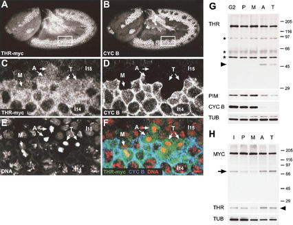
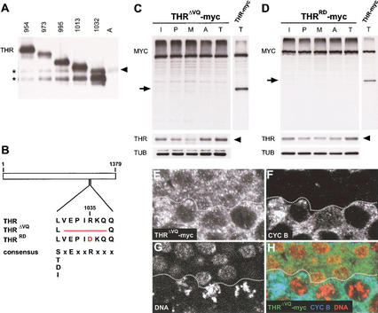
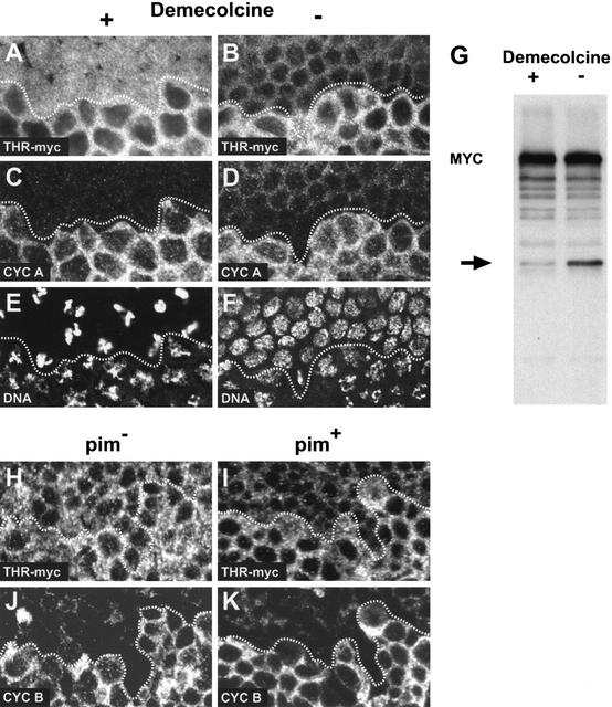
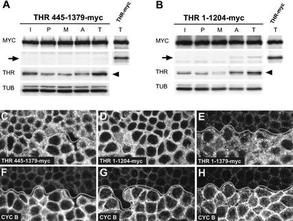
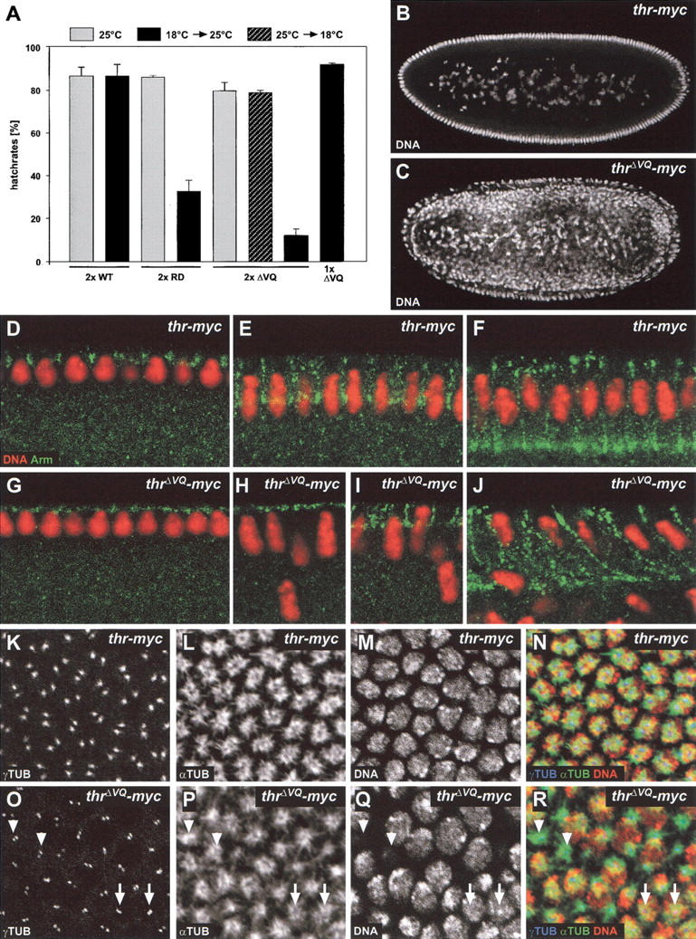
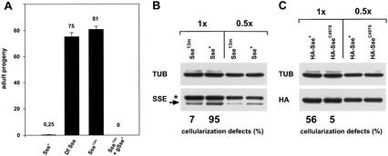
Sister-chromatid separation in mitosis requires proteolytic cleavage of a cohesin subunit. Separase, the corresponding protease, is activated at the metaphase-to-anaphase transition. Activation involves proteolysis of an inhibitory subunit, securin, following ubiquitination mediated by the anaphase-promoting complex/cyclosome. In Drosophila, the securin PIM associates not only with separase (SSE), but also with an additional protein, THR. Here we show that THR is cleaved after the metaphase-to-anaphase transition. THR cleavage only occurs in functional SSE complexes and in a region that matches the separase cleavage-site consensus. Mutations in this region abolish mitotic THR cleavage. These results indicate that THR is cleaved by SSE. Expression of noncleavable THR variants results in cold-sensitive maternal-effect lethality. This lethality can be suppressed by a reduction of catalytically active SSE levels, indicating that THR cleavage inactivates SSE complexes. THR cleavage is particularly important during the process of cellularization, which follows completion of the last syncytial mitosis of early embryogenesis, suggesting that Drosophila separase has other targets in addition to cohesin subunits.


Figures

Figure 1
Figure 1
THR is degraded during mitosis. Embryos (A,B) expressing gthr–myc were fixed at the stage of mitosis 14 and labeled with antibodies against the myc epitope (A,C,F), cyclin B (B,D,F), and a DNA stain (E,F). The boxed area in A and B is shown in CF. Red, green, and blue in the merged panel F represent DNA, anti-myc, and anti-cyclin B labeling, respectively. M, metaphase; A, anaphase; T, telophase; I14, interphase 14; I15, interphase 15. (G) Synchronous progression through mitosis 14 was induced, and extracts were prepared from embryos with all cells in G2 before mitosis 14 (G2), as well as in prophase (P), metaphase (M), anaphase (A), and telophase (T) of mitosis 14. Extracts were analyzed by immunoblotting using antibodies against THR (THR), PIM (PIM), cyclin B (CYC B), and tubulin (TUB). A 47-kD fragment appearing after the metaphase-to-anaphase transition is indicated by an arrowhead. Asterisks indicate cross-reacting bands. (H) Extracts from gthr–myc embryos during interphase (I), prophase (P), metaphase (M), anaphase (A), and telophase (T) of the synchronous syncytial blastoderm cycles were analyzed by immunoblotting using antibodies against the myc epitope (MYC), THR (THR), and tubulin (TUB). Mitotic cleavage products of THR–myc and endogenous THR are indicated by an arrow and an arrowhead, respectively.
Figure 2
Figure 2
Mapping the mitotic THR cleavage site. (A) C-terminal THR fragments were generated in vitro and resolved next to an anaphase embryo extract (lane A, same extract as in Fig. 1G, lane A). THR fragments were detected by immunoblotting with anti-THR antibodies. The numbers above the lanes indicate the amino acid position at which the C-terminal THR fragments start. The arrowhead indicates the C-terminal THR fragment generated in vivo after the metaphase-to-anaphase transition. Asterisks indicate partial products of the THR fragments generated in vitro. (B) Schematic illustration of the mitotic cleavage region within THR, THRΔVQ, and THRRD. In addition, the separase cleavage-site consensus sequence (Hauf et al. 2001) is shown below the THR sequences. Cleavage by separase occurs C-terminal from the conserved arginine residue. (C,D) Extracts from gthrΔVQ–myc (C) or gthrRD–myc (D) embryos during interphase (I), prophase (P), metaphase (M), anaphase (A), and telophase (T) of the synchronous syncytial blastoderm cycles were analyzed by immunoblotting using antibodies against the myc epitope (MYC), THR (THR), and tubulin (TUB). In addition, a telophase extract from gthr–myc embryos was analyzed in parallel (right lanes). Mitotic cleavage products of THR–myc and endogenous THR are indicated by arrows and arrowheads, respectively. (EH) Embryos expressing gthrΔVQ–myc were fixed at the stage of mitosis 14 and labeled with antibodies against the myc epitope (E), cyclin B (F), and a DNA stain (G). Red, green, and blue in the merged image (H) represent labeling of DNA, myc, and cyclin B, respectively. The epidermal region shown corresponds to the boxed region in Figure 1A. Cells below the dotted line are in G2 before mitosis 14, whereas cells above the dotted line have progressed through mitosis 14 and are mostly in early interphase of cycle 15. Note that THRΔVQ–myc is still present at high levels in these cells, in contrast to THR–myc (see Fig. 1C–F).
Figure 3
Figure 3
THR cleavage is inhibited in cells arrested by the mitotic spindle checkpoint and in pim mutants. (AF) Embryos expressing gthr–myc were permeabilized and incubated in the presence (A,C,E) or absence (B,D,F) of the microtubule inhibitor demecolcine while progressing through mitosis 14. After fixation, embryos were labeled with antibodies against the myc epitope (A,B), cyclin A (C,D), and a DNA stain (E,F). Comparable epidermal regions are shown. Cells below the dotted line are in G2 before mitosis 14, whereas cells above the dotted line have progressed into mitosis 14 and are arrested with condensed chromatin (E) or are already in early interphase of cycle 15 (F). THR–myc is present at high levels in arrested cells (A), and only at low levels in interphase 15 cells (B). (G) gthr–myc embryos during the syncytial blastoderm cycles were permeabilized and incubated in the presence (+) or absence (−) of demecolcine. Embryo extracts were analyzed by immunoblotting with anti-myc. The THR–myc fragment appearing after the metaphase-to-anaphase transition is indicated by an arrow. (HK) pim embryos (H,J) and pim+ sibling embryos (I,K) expressing gthr–myc were fixed at the stage of mitosis 15 and labeled with antibodies against the myc epitope (H,I) and cyclin B (J,K). In the epidermal region shown, cells below the dotted line are in G2 before mitosis 15, whereas cells above the dotted line have progressed through mitosis 15 and are mostly in early interphase of cycle 16. These latter cells have high levels of THR–myc in pim embryos (H), and only low levels in pim+ sibling embryos (I).
Figure 4
Figure 4
THR cleavage occurs only within functional SSE complexes. (A,B) Extracts from gthr 445–1379–myc (A) or gthr 1–1204–myc (B) embryos during interphase (I), prophase (P), metaphase (M), anaphase (A), and telophase (T) of the synchronous syncytial blastoderm cycles were analyzed by immunoblotting using antibodies against the myc epitope (MYC), THR (THR), and tubulin (TUB). In addition, a telophase extract from gthr–myc embryos was analyzed in parallel (right lanes). The proteins THR 445–1379–myc, THR 1–1204–myc, and THR–myc all contain the cleavage region and associate with SSE. Mitotic cleavage products of THR–myc and endogenous THR are indicated by arrows and arrowheads, respectively. (CH) Embryos expressing gthr 445–1379–myc (C,F), gthr 1–1204–myc (D,G), or gthr–myc (E,H) were fixed at the stage of mitosis 14 and labeled with antibodies against the myc epitope (CE) and cyclin B (FH). Comparable epidermal regions are shown. Cells below the dotted lines are in G2 before mitosis 14, whereas cells above the dotted lines have progressed through mitosis 14 and are in early interphase of cycle 15. Note that THR 445–1379–myc (C) and THR 1–1204–myc (D) are still present at high levels in these cells, in contrast to THR–myc (E).
Figure 5
Figure 5
Phenotype associated with expression of noncleavable THR. (A) Noncleavable THR present during early embryonic development causes cold-sensitive, maternal-effect lethality. Eggs were collected at 25°C for 1 h from females homozygous for the transgene insertions gthr–myc (2× WT), gthrRD–myc (2× RD), or gthrΔVQ–myc (2× ΔVQ), or heterozygous for gthrΔVQ–myc (1× ΔVQ). Eggs were incubated at 25°C (gray bars); or shifted to 18°C after 3.5 h (hatched bar); or shifted to 18°C for 4.5 h, followed by a shift back to 25°C (black bars). The larval hatch rates (% of hatched eggs) are given as average values obtained from three independent experiments. (B,C) Noncleavable THR causes internalization of nuclei during early embryonic development. Embryos derived from females homozygous for gthr–myc (B) or gthrΔVQ–myc (C) were incubated during their early development at 18°C, fixed, and stained for DNA. (DJ) Cellularization is delayed in THRΔVQ embryos. Cellularizing THR (DF) and THRΔVQ (GJ) embryos were fixed and labeled with an antibody against Armadillo (Arm, green) and a DNA stain (DNA, red). (KR) Noncleavable THR affects centrosome separation. THR (KN) or THRΔVQ (OR) embryos were fixed during cellularization at 18°C, stained for DNA, and labeled with antibodies against γ-tubulin (γTUB) and α-tubulin (αTUB). Apical confocal sections that contained the γTUB signals were stacked (K,L,O,P), and a lower section was taken for DNA (M,Q). Arrows indicate unseparated centrosomes. Arrowheads denote positions where nuclei had dropped into the interior of the embryo and had left behind centrosomes and microtubule asters. (N) Merge of K, L, and M; (R) merge of O, P, and Q. Red, green, and blue in the merged images indicate labeling of DNA, αTUB, and γTUB, respectively.
Figure 6
Figure 6
THR cleavage limits SSE activity. (A) Females carrying two copies of the gthrΔVQ–myc transgene in a genetic background that was Sse+/Sse+ (Sse+), Df(3L)SseA/Sse+ (Df Sse), Sse13m/Sse+ (Sse13m), or Sse13m, gSse+/Sse+ (Sse13m + gSse+) were crossed to w1 males. Df(3L)SseA deletes Sse; Sse13m is a null allele and gSse+ is a transgene constructed with a genomic fragment providing Sse+ function (Jäger et al. 2001). Progeny developing at 18°C were counted. Average values of progeny/day and females obtained from at least four independent experiments are given for each cross. (B,C) Females carrying two copies of the gthrΔVQ–myc transgene in a genetic background that was either Sse+/Sse+ (Sse+) or Sse13m/Sse+ (Sse13m) were crossed to w1 males (B). Females that carried two copies of the gthrΔVQ–myc transgene in an Sse13m/Sse+ genetic background and in addition expressed HA–Sse+ (HA–Sse+) or HA–SseC497S (HA–SseC497S) were also crossed to w1 males (C). HA–SseC497S encodes a catalytically inactive SSE mutant. Embryos from these crosses were used to quantitate cellularization defects at 18°C and to prepare protein extracts for immunoblotting. Extracts were loaded either undiluted (1×) or in a 1:2 dilution (0.5×). Blots were probed with antibodies against SSE (SSE; arrow in B), the HA epitope (HA; C) or tubulin (TUB) as a loading control. The asterisk in B indicates a cross-reacting band.

Similar articles

Cited by

References

    1. Alexandru G, Uhlmann F, Mechtler K, Poupart MA, Nasmyth K. Phosphorylation of the cohesin subunit Scc1 by Polo/Cdc5 kinase regulates sister chromatid separation in yeast. Cell. 2001;105:459–472. - PubMed
    1. Brinkley BR, Cartwright J., Jr Cold-labile and cold-stable microtubules in the mitotic spindle of mammalian cells. Ann NY Acad Sci. 1975;253:428–439. - PubMed
    1. Chen L, Puri R, Lefkowitz EJ, Kakar SS. Identification of the human pituitary tumor transforming gene (hPTTG) family: Molecular structure, expression, and chromosomal localization. Gene. 2000;248:41–50. - PubMed
    1. Ciosk R, Zachariae W, Michaelis C, Shevchenko A, Mann M, Nasmyth K. An ESP1/PDS1 complex regulates loss of sister chromatid cohesion at the metaphase to anaphase transition in yeast. Cell. 1998;93:1067–1076. - PubMed
    1. Clarkson M, Saint R. A His2AvDGFP fusion gene complements a lethal His2AvD mutant allele and provides an in vivo marker for Drosophila chromosome behavior. DNA Cell Biol. 1999;18:457–462. - PubMed

Publication types

MeSH terms