. 2002 Sep 15;543(Pt 3):871-87.
doi: 10.1113/jphysiol.2002.021915.

Loss of enteric motor neurotransmission in the gastric fundus of Sl/Sl(d) mice

Affiliations

Loss of enteric motor neurotransmission in the gastric fundus of Sl/Sl(d) mice

Elizabeth A H Beckett et al. J Physiol. .

Abstract

Studies of W/W(V) mice, which lack intramuscular interstitial cells of Cajal (IC-IM), have suggested that IC-IM act as mediators of enteric motor neurotransmission in the gastrointestinal tract. We have studied Sl/Sl(d) mice, which lack the ability to make membrane-bound stem cell factor, to determine the consequences of inappropriate stem cell factor expression on IC-IM populations and on enteric motor neurotransmission. IC-IM were found within the circular and longitudinal muscles of the gastric fundus of wild-type mice. IC-IM were intimately associated with motor nerve terminals and nerve varicosities formed synaptic structures with these cells. IC-IM were also connected with neighbouring smooth muscle cells via gap junctions. Immunohistochemistry and electron microscopy showed that IC-IM were absent from fundus muscles of Sl/Sl(d) mice, but the density of excitatory and inhibitory nerves was not significantly different than in wild-type muscles. Loss of IC-IM was associated with decreased membrane noise (unitary potentials) and significant reductions in post-junctional excitatory and inhibitory enteric nerve responses. Reductions in neural responses were not due to defects in smooth muscle cells as responses to exogenous ACh and K(+)-induced depolarization were normal in Sl/Sl(d) mice. Responses to neurally released ACh were revealed in Sl/Sl(d) mice by inhibiting ACh breakdown with the acetylcholinesterase inhibitor neostigmine. Inhibitory nerve stimulation elicited inhibitory junction potentials (IJPs) and relaxations in wild-type mice. IJPs were reduced in amplitude and relaxation responses were absent in Sl/Sl(d) mice. These observations suggest that membrane-bound stem cell factor is essential for development of IC-IM and that the close, synaptic-like relationship between nerve terminals and IC-IM may be the primary site of innervation by enteric motor neurons in gastric muscles.


Figures

Figure 1
Figure 1. Distribution of IC-IM within the circular and longitudinal muscle layers of wild-type and Sl/Sld murine gastric fundus muscles
A, Kit-like immunopositive IC-IM within the circular (arrows) and longitudinal muscle layers (arrowheads) of a wild-type control animal. IC-IM are elongate spindle shaped cells that run parallel to the muscle fibres of each layer. Kit-like immunohistochemistry revealed an absence of IC-IM in the circular and longitudinal muscle layers of age-matched Sl/Sld mutant siblings (B). Scale bar shown in B also applies to A.
Figure 2
Figure 2. Confocal microscopy revealing the relationship between enteric motor neurons and ICC within the circular muscle layer (IC-IM)
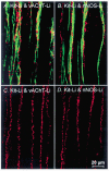
A, an overlay of confocal fluorescence micrographs showing Kit-like immunopositive IC-IM (Kit-Li; green) and vesicular acetylcholine transporter-like immunoreactivity (vAChT-Li; red) to label excitatory motor neurons within the circular muscle layer of wild-type fundus. Note the close anatomical relationship between excitatory enteric nerves and IC-IM. B, an overlay of fluorescence micrographs showing the close association between IC-IM (Kit-Li; green) and neuronal nitric oxide synthase-like immunoreactivity (nNOS-Li; red) to label inhibitory motor neurons in wild-type fundus. Note the close apposition of IC-IM with varicose terminals of inhibitory motor neurons. C and D are from an Sl/Sld animal. Despite the absence of Kit-Li IC-IM in the circular muscle layer of Sl/Sld mutants, both vAChT-Li and nNOS-Li enteric motor nerves (red) are distributed in a manner similar to that of controls (C and D, respectively). Images shown are digital reconstructions taken from 3 × 0.56 μm scans. Scale bar shown in D is for all panels.
Figure 3
Figure 3. Transmission electron microscopy reveals the morphology of IC-IM and their ultrastructural relationship with enteric motor nerve endings
A, an IC-IM (IC) within the circular muscle layer interposed between nerve processes (N) and circular smooth muscle cells (CM). The cytoplasm of IC-IM were characteristically electron dense compared to smooth muscle cells and possessed an abundance of mitochondria (B; frame in B is magnified in C). Free ribosomes, a prominent Golgi apparatus and a well-developed endoplasmic reticulum were also observed within the cytoplasm of IC-IM (A, D and E). A discontinuous basal lamina was associated with the plasma membrane that distinguished these cells from macrophages or fibroblasts, and the lack of dense bodies and contractile filaments distinguish the IC-IM from smooth muscle cells (E and F). Varicose nerve fibre endings possessed large dense cored and smaller electron-lucent vesicles and formed morphological appositions with IC-IM of distances < 10 nm. Close contacts (arrowheads) and synaptic-like specializations (arrows) appeared to exist between the membranes of enteric nerve varicosities and IC-IM (C and D; inset in D is a higher power image of the contact indicated by the left-hand arrow). IC-IM formed gap junctions with one another (arrow in E and inset) and with neighbouring smooth muscle cells (arrowhead in E, and arrow in F; shown at greater magnification in inset). IC-IM within the longitudinal muscle layer had a similar ultrastructural appearance to IC-IM in the circular layer and also formed close associations with enteric nerves, adjacent IC-IM and neighbouring smooth muscle cells (B and C). Cells with the ultrastructual features of IC-IM were absent in Sl/Sld mutants (G). Enteric nerves of Sl/Sld mutants (N) contained both large dense cored and smaller electron-lucent vesicles, but these nerves never formed the synaptic-like membrane specializations with smooth muscle cells (CM) (inset in G, arrowheads).
Figure 4
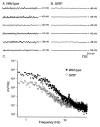
Figure 4. Intracellular recordings of wild-type and Sl/Sld fundus muscles reveals differences in the basal resting membrane potential
A, the typical activity recorded from six wild-type animals (20 s traces) revealing membrane potential fluctuations. B shows recordings from six age-matched Sl/Sld mutant siblings. The membrane potential fluctuations observed in wild-type controls were greatly reduced in these tissues. C shows differences in power spectral densities of the recordings from wild-type (•) and Sl/Sld mutants (○). Membrane fluctuations of wild-type fundus muscles had greater power in the 0.3–10 Hz range compared to Sl/Sld mutants.
Figure 5
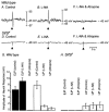
Figure 5. Differences in neural responses to electrical field stimulation (EFS) in wild-type and Sl/Sld mutant animals
EFS (arrow; 0.5 ms pulse at supramaximal voltage) produced a bi-phasic electrical response in wild-type muscles, characterized by a rapid excitatory junction potential (EJP) that was followed by an inhibitory junction potential (IJP; A). Nω-Nitro-l-arginine (l-NA; 100 μM) reduced the amplitude of IJPs and potentiated the amplitudes of the EJPs (B). After addition of l-NA, atropine (1 μM) completely blocked the EJPs, providing evidence that the EJP response was cholinergic in nature. In Sl/Sld mutant animals, EJPs were absent and IJPs were greatly reduced in amplitude (D). l-NA had little or no effect on EJPs and failed to reduce the attenuated IJPs (E). In the presence of l-NA, atropine had no effect (F). The effects of l-NA and atropine on EJPs and IJPs on 10 wild-type (▪) and 10 Sl/Sld mutant animals (□) is summarized in G and H, respectively.
Figure 6
Figure 6. Multiple pulses of EFS (1–20 Hz; 0.5 ms duration for 1 s) did not reveal significant excitatory or inhibitory responses in Sl/Sld mutants
A shows neural responses of a wild-type animal to EFS (1 pulse, 5 and 10 Hz for 1 s) delivered at the time indicated by the arrow and bars. B shows the neural responses of an Sl/Sld mutant to similar EFS parameters. C and D show the summarized data for EJP (C) and IJP responses (D) to EFS recorded from wild-type and Sl/Sld mutants before (circles; filled for wild-type and open for Sl/Sld) and after (squares; filled for wild-type and open for Sl/Sld) the addition of l-NA (100 μM).
Figure 7
Figure 7. EFS revealed differences in the mechanical responses to nerve stimulation of wild-type and Sl/Sld mutant muscles
A shows mechanical responses to EFS (5 Hz and 10 Hz of EFS, 0.5 ms duration for 30 s delivered at the time indicated by the bars using supramaximal voltage) of wild-type fundus muscle strips. l-NA (100 μM) abolished relaxations induced by EFS and unmasked large contractile responses (B). In the continued presence of l-NA, atropine (1 μM) significantly attenuated the amplitude of contractions in response to EFS (C). D shows mechanical responses of an Sl/Sld mutant in control conditions using the same EFS stimulus parameters. In Sl/Sld muscle strips relaxations were not evoked and contractions induced by EFS were significantly smaller compared to wild-type animals. l-NA (100 μM) and atropine (1 μM) had little effect on the amplitude of Sl/Sld fundus muscle contractions (E and F). G shows a summary of the data of wild-type muscles in the presence of l-NA to reveal excitatory responses, before (•) and after (▪) the addition of atropine. A significant portion of the contractile responses of wild-type muscles was blocked by atropine. H shows a summary of the data from Sl/Sld muscles. Contractions evoked in Sl/Sld muscles in control conditions (▿) were enhanced by l-NA (○) but remained significantly smaller in amplitude than those evoked in wild-type muscles in identical conditions. Sl/Sld muscle responses in the presence of l-NA and atropine (□) were similar in amplitude to wild-type muscle responses in these conditions.
Figure 8
Figure 8. Electrical and mechanical responses of wild-type and Sl/Sld muscles to exogenous acetylcholine (ACh) and sodium nitroprusside (SNP)
ACh (1.0 μM) depolarized muscles from wild-type and Sl/Sld mutants in a similar manner. A and B show depolarization responses of wild-type and Sl/Sld muscles to ACh (1.0 μM, arrows), respectively. C is the summarized data for depolarizations to ACh in wild-type (▪) and Sl/Sld mutants (□). Summarized data were not statistically significant between wild-type and Sl/Sld mutants. D and E show the contractile responses of wild-type and Sl/Sld muscles to ACh (1.0 μM, arrows). F shows the summarized contractile responses for wild-type (▪) and Sl/Sld mutants (□) in response to acetylcholine. Contractile responses were not statistically different between the two groups. G and H show hyperpolarization responses of wild-type and Sl/Sld muscles in response to addition of the nitric oxide donor sodium nitroprusside (SNP, 1 μM, arrows), respectively. Wild-type muscles hyperpolarized following addition of SNP but Sl/Sld mutants showed little or no response (H shows one of the five muscle strips, from a total of six, hyperpolarized slightly in response to SNP). Summarized data of the hyperpolarization responses of wild-type (▪) and Sl/Sld mutants (□) are shown in I. * Significant difference (P < 0.001) between wild-type and Sl/Sld mutants in response to SNP. J and K show relaxations in response to SNP (1 μM) for wild-type and Sl/Sld mutants, respectively. No statistical difference was observed in the relaxation responses between these two groups (L).
Figure 9
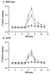
Figure 9. [14C]Choline release from wild-type and Sl/Sld muscles in response to EFS
Tissues were stimulated using EFS at 40 min intervals for 1 min (5 Hz, 0.3 ms duration pulses; 60 V) and [14C]choline overflow was assayed from superfusion samples. [14C]Choline overflow is plotted as a percentage of fractional release with time of collection. A shows a summary of [14C]choline overflow, plotted as a percentage of total [14C]choline from wild-type fundus muscles (n = 10), in response to EFS under control conditions (○). [14C]Choline overflow in response to EFS was inhibited by TTX (1 μM, •) and recovered after washout of TTX (□). B shows a summary of [14C]choline overflow from Sl/Sld mutant tissues (n = 10) in response to EFS under control conditions (○). This release was also inhibited by TTX (1 μM, •) and recovered after washout (□).
Figure 10
Figure 10. Inhibition of endogenous acetylcholinesterase with neostigmine potentiated the fast EJP and revealed a slowly developing membrane depolarization in wild-type tissues and revealed a slowly developing depolarization in Sl/Sld animals
A shows the electrical responses of wild-type muscles to EFS (single pulses 0.4 ms duration, supramaximal voltage, delivered at time indicated by arrow). B shows responses to EFS after the addition of l-NA (100 μM). l-NA greatly reduced or abolished electrical responses at all stimulus durations. C shows recordings in the presence of l-NA and the acetylcholinesterase inhibitor neostigmine (Neo, 1 μM). Neostigmine produced a slowly developing membrane depolarization at all pulse durations tested. Both fast EJPs and slowly developing depolarizations were completely blocked by atropine (D) at all pulse durations, suggesting that muscarinic receptors mediated these responses. Electrical recordings in A-D were all from the same cell. E shows the lack of response to Sl/Sld muscles to single pulse (0.4 ms duration) EFS. After the addition of l-NA (100 μM) Sl/Sld muscles still showed no responses to EFS (F). In the presence of l-NA and neostigmine a slowly developing membrane depolarization occurred in response to EFS (G) that was completely inhibited by the addition of atropine (1 μM, H). Electrical recordings in E-H were all from the same cell.

Similar articles

Cited by

References

    1. Alberts P, Bartfai T, Stjarne L. The effects of atropine on [3H]-acetylcholine secretion from guinea-pig myenteric plexus evoked electrically or by high potassium. Journal of Physiology. 1982;329:93–112. - PMC - PubMed
    1. Brannan CI, Lyman SD, Williams DE, Eisenman J, Anderson DM, Cosman D, Bedell MA, Jenkins NA, Copeland NG. Steel-Dickie mutation encodes a c-kit ligand lacking transmembrane and cytoplasmic domains. Proceedings of the National Academy of Sciences of the USA. 1991;88:4671–4674. - PMC - PubMed
    1. Burns AJ, Lomax AE, Torihashi S, Sanders KM, Ward SM. Interstitial cells of Cajal mediate inhibitory neurotransmission in the stomach. Proceedings of the National Academy of Sciences of the USA. 1996;93:12008–12013. - PMC - PubMed
    1. Daniel EE, Posey-Daniel V. Neuromuscular structures in opossum esophagus: role of interstitial cells of Cajal. American Journal of Physiology. 1984;246:G305–315. - PubMed
    1. Desai KM, Sessa WC, Vane JR. Involvement of nitric oxide in the reflex relaxation of the stomach to accommodate food or fluid. Nature. 1991;351:477–479. - PubMed

Publication types

MeSH terms

LinkOut - more resources