. 2002 Sep 15;543(Pt 3):933-45.
doi: 10.1113/jphysiol.2001.015750.

A possible dual site of action for carbon monoxide-mediated chemoexcitation in the rat carotid body

Affiliations

A possible dual site of action for carbon monoxide-mediated chemoexcitation in the rat carotid body

C Barbé et al. J Physiol. .

Abstract

High tensions of carbon monoxide (CO), relative to oxygen, were used as a tool to investigate the mechanism of chemotransduction. In an in vitro whole organ, rat carotid body preparation, CO increased sinus nerve chemoafferent discharge in the dark, an effect that was significantly reduced (by ca 70 %) by bright white light and by the removal of extracellular Ca(2+) from the superfusate or by the addition of either Ni(2+) (2 mM) or methoxyverapamil (100 microM). Addition of the P(2) purinoceptor antagonist pyridoxalphosphate-6-azophenyl-2',4'-disulphonic acid (50 microM) also significantly reduced the neural response to CO. In perforated patch, whole-cell recordings of isolated rat type I cells, CO induced a depolarisation of ca 11 mV and a decrease in the amplitude of an outward current around and above the resting membrane potential. Membrane conductance between -50 and -60 mV was significantly reduced by ca 40 % by CO. These effects were not photolabile and were present also when a 'blocking solution' containing TEA, 4-AP, Ni(2+) and zero extracellular Ca(2+) was used. In conventional whole-cell recordings, CO only decreased current amplitudes above +10 mV and was without effect around the resting membrane potential. These data demonstrate a direct effect of CO upon type I cell K(+) conductances and strongly suggest an effect upon a background, leak conductance that requires an intracellular mediator. The photolabile effect of CO only upon afferent neural discharge adds further evidence to a dual site of action of CO with a separate action at the afferent nerve terminal that, additionally, requires the permissive action of the neurotransmitter ATP.


Figures

Figure 1
Figure 1. Carbon monoxide sensitivity is light- and extracellular [Ca2+] dependent
Single fibre chemoreceptor discharge was recorded and binned into 10 s periods. CO (320 mmHg) was delivered as shown (horizontal bars), initially in the presence of bright white light. After a period of at least 2 min, the light was switched off (light off; horizontal thick bars) for a brief period. On the left can be seen the normal, brisk and intense control response to CO with normal extracellular Ca2+. On the right, in the absence of extracellular Ca2+ (0 Ca; horizontal thin bar), CO was without effect when the light was switched off. Inset shows eight superimposed afferent action potentials from the single fibre recording with scale bars representing 1 mV and 1 ms.
Figure 2
Figure 2. Effect of ATP receptor antagonism upon carbon monoxide sensitivity
Single fibre chemoreceptor afferent discharge was recorded and binned into 10 s periods and steady-state discharge averaged over at least 30 s. All recordings were made in the absence of light. CO (320 mmHg)-stimulated discharge was inhibited by the addition of the ATP receptor antagonist PPADS (50 μM). The inhibitory effect of PPADS was reversed by washout for 30–60 min. Data are given as means ± s.e.m.*P < 0.05, n = 8.
Figure 3
Figure 3. Carbon monoxide depolarises isolated type I cells
Example of the effect of acidosis and CO in one cell on the resting membrane potential. The recording was made during current clamp using the amphotericin B, whole-cell configuration. Acidosis (PCO2 = 76 mmHg; PO2 = 60 mmHg) and CO (PCO = 180 mmHg; PO2 = 60 mmHg) both reversibly depolarised the type I cell. Experiments were performed in the absence of light.
Figure 4
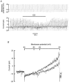
Figure 4. Carbon monoxide decreases the amplitude of outward currents above -50 mV
A, an example of outward currents obtained during 300 ms pulses to membrane potentials between -90 and +30 mV from a holding potential of -70 mV, in control conditions (left) and during perfusion with CO (right). Recordings were made using the amphotericin B, whole-cell configuration. B, the mean ± s.e.m. (n = 7) current density-voltage relationships in control conditions (•), in the presence of CO (○) and after recovery from CO (⋆). The inset shows an expanded scale of the same current density-voltage relationships to highlight the effect of CO at membrane potentials where current density was around 0 pA pF−1. C, the mean ± s.e.m. (n = 7) of the CO-sensitive component of the total current density-voltage relationship determined as the difference between the current density amplitudes in control conditions and in the presence of CO. Experiments were performed in the absence of light.
Figure 5
Figure 5. Carbon monoxide decreases the amplitude of outward currents above +10 mV
A, example of outward currents obtained during 300 ms pulses to membrane potentials between -90 and +30 mV from a holding potential of -70 mV, in control conditions (left) and during perfusion with CO (right). Recordings were made using the conventional, whole-cell configuration. B, the mean ± s.e.m. (n = 7) current density-voltage relationships in control conditions (•), in the presence of CO (○) and after recovery from CO (⋆). The inset shows an expanded scale of the same current density-voltage relationships to highlight the negligible effect of CO at membrane potentials where current density was around 0 pA pF−1. C, the mean ± s.e.m. (n = 7) of the CO-sensitive component of the total current density-voltage relationship determined as the difference between the current density amplitudes in control conditions and in the presence of CO. Experiments were performed in the absence of light.
Figure 6
Figure 6. Carbon monoxide decreases resting membrane conductance
Cells were voltage clamped at -70 mV and subjected to a 2 s voltage ramp from -90 to -30 mV every 6 s. A, voltage protocol and resultant currents during a single experiment. During CO exposure there was a reversible decrease in the amplitude of the current. B, mean current responses to voltage ramps. The lower trace is that during CO exposure. Slope conductances were measured between -50 and -60 mV. Recordings were made using the amphotericin B, whole-cell configuration. Experiments were performed in the absence of light.
Figure 7
Figure 7. Carbon monoxide decreases a K+ resting membrane conductance
A, average current-voltage relationships of the CO-sensitive current in the presence of K+ channel blockers, Ni2+ and the absence of Ca2+ with 4.5 mm K+ (•, n = 4) or with 20 mm K+ (○, n = 5). Note the change of the reversal potential between these two different conditions. B, summary mean bar graph of the reversal potential obtained in control conditions (4.5 mm K+, n = 7), in the presence of K+ channel blockers, Ni2+ and the absence of Ca2+ (4.5 mm K+ + blockers, n = 4) and in this last condition but with 20 mm K+ (20 mm K+ + blockers, n = 5). C, plot of the measured shift in reversal potential observed following the increase in K+ (Na+ replacement, •).The open circles indicate calculated shifts in reversal potential expected for a purely K+-selective conductance. Data represent means ± s.e.m. in 4–5 cells. Recordings were made using the amphotericin B, whole-cell configuration and performed in the absence of light.

Similar articles

Cited by

References

    1. Al-Hashem F, Barbe C, Kumar P. Interaction of ATP and CO2 upon carotid body chemosensitivity in vitro. Journal of Physiology. 2001;536.P:142P.
    1. Al-Hashem F, Conway AF, Kumar P. Calcium- and age-dependent effects of carbon monoxide upon chemodischarge in the rat carotid body in vitro. Journal of Physiology. 2000;523.P:296P.
    1. Anichkov SV, Belen'kii ML. Pharmacology of the Carotid Body Chemoreceptors. New York: Macmillan; 1963.
    1. Barbé C, Dubuis E, Vandier C, Kumar P. Effect of carbon monoxide on whole-cell currents of the isolated rat carotid body type I cell. Journal of Physiology. 2001;533.P:108–109P.
    1. Buckler KJ. A novel oxygen-sensitive potassium current in rat carotid body type I cells. Journal of Physiology. 1997;498:649–662. - PMC - PubMed

Publication types

MeSH terms

LinkOut - more resources