. 2002 Sep 23;87(7):805-12.
doi: 10.1038/sj.bjc.6600547.

Evaluation of 2-deoxy-D-glucose as a chemotherapeutic agent: mechanism of cell death

Affiliations

Evaluation of 2-deoxy-D-glucose as a chemotherapeutic agent: mechanism of cell death

R L Aft et al. Br J Cancer. .

Abstract

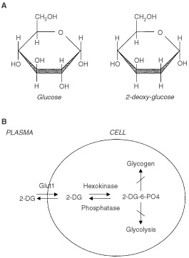
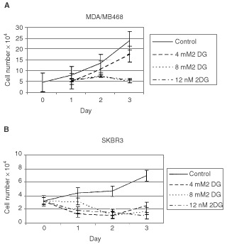
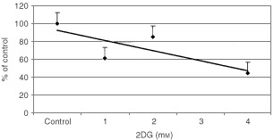
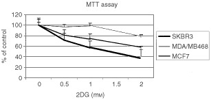
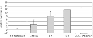
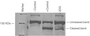
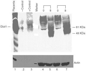
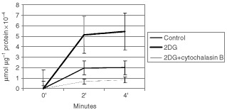
Nutrient deprivation has been shown to cause cancer cell death. To exploit nutrient deprivation as anti-cancer therapy, we investigated the effects of the anti-metabolite 2-deoxy-D-glucose on breast cancer cells in vitro. This compound has been shown to inhibit glucose metabolism. Treatment of human breast cancer cell lines with 2-deoxy-D-glucose results in cessation of cell growth in a dose dependent manner. Cell viability as measured by the 3-(4,5-dimethylthiazol-2-yl)-2,5-diphenyl tetrazolium bromide conversion assay and clonogenic survival are decreased with 2-deoxy-D-glucose treatment indicating that 2-deoxy-D-glucose causes breast cancer cell death. The cell death induced by 2-deoxy-D-glucose was found to be due to apoptosis as demonstrated by induction of caspase 3 activity and cleavage of poly (ADP-ribose) polymerase. Breast cancer cells treated with 2-deoxy-D-glucose express higher levels of Glut1 transporter protein as measured by Western blot analysis and have increased glucose uptake compared to non-treated breast cancer cells. From these results we conclude that 2-deoxy-D-glucose treatment causes death in human breast cancer cell lines by the activation of the apoptotic pathway. Our data suggest that breast cancer cells treated with 2-deoxy-D-glucose accelerate their own demise by initially expressing high levels of glucose transporter protein, which allows increased uptake of 2-deoxy-D-glucose, and subsequent induction of cell death. These data support the targeting of glucose metabolism as a site for chemotherapeutic intervention by agents such as 2-deoxy-D-glucose.


Figures

Figure 1
Figure 1
(A) Structural comparison of glucose and 2-deoxy-D-glucose. 2DG and glucose differ at the second carbon. (B) Schematic diagram of 2-DG action. 2DG enters the cell through the glucose transporter and is phosphorylated by hexokinase. Due to low levels of intracellular phosphatase, 2-DG-PO4 is trapped in the cell. 2-DG-PO4 is unable to undergo further metabolism. High intracellular levels of 2-DG-6-PO4 cause allosteric and competitive inhibition of hexokinase. This results in inhibition of glucose metabolism.
Figure 2
Figure 2
Effect of 2-DG on growth of breast cancer cell lines. SkBr3 and MDA/MB468 breast cancer cell lines were grown in the absence or presence of various concentrations of 2DG. 2DG was added on day zero and the media was not changed during the duration of the experiment. Cell number was determined daily. Each point represents triplicate cultures.
Figure 3
Figure 3
Clonogenic survival after 2DG treatment. SkBr3 cells were treated with varying concentrations of 2DG for 4 h and replated at low density. Colonies were counted 14 days after plating and those containing 25 cells or greater were scored as positive. Each point represents triplicate cultures.
Figure 4
Figure 4
MTT conversion after 2DG treatment. SkBr3, MDA/MB468, and MCF7 cells were incubated with the indicated concentrations of 2DG for 4 h. MTT was added the amount of reduced formazan product determined spectrophotometrically. Results are expressed as per cent of control non-treated cells.
Figure 5
Figure 5
Caspase 3 activation in SkBr3 cells treated with 2DG. SkBr3 cells were treated with 16 mM 2DG for the 4 or 6 h. Cells were harvested and caspase 3 activity measured using a spectrophotometric assay according to the manufacturer's instructions. The caspase 3 inhibitor DEVD-FMK was added 30 min prior to the addition of substrate in cells which had been incubated with 16 mM 2DG for 6 h. Each point represents the average of triplicate cultures. Control cells were incubated for 6 h in the presence of media only.
Figure 6
Figure 6
PARP activation by SkBr3 cells treated with 2-DG. Cells were treated for 6 h with 12 mM 2DG or 2 μM staurosporine (+control). Western blot analysis was performed using a monoclonal antibody to PARP. Normal (uncleaved) and the PARP cleavage product are indicated.
Figure 7
Figure 7
Glut-1 transporter protein levels in breast cancer cells lines treated with 2-DG. SkBr3 breast cancer cells were treated with 2DG for 48 or 72 h with 8 mM 2DG (8) or without 2DG (0). Protein was isolated, size separated on a 10% polyacrylamide gel. A Western blot was performed using a polyclonal anti-Glut-1 antibody. Positive controls are protein isolated from human placenta and membranes isolated from Glut1 injected Xenopus oocytes (+control). Sham injected oocytes were used as a negative control (−control). Glut1 protein is indicated. Molecular weight markers are labelled. The protein blot was reacted with anti-actin antibody after removal of the Glut1 antibody (lower panel).
Figure 8
Figure 8
Glucose uptake by SkBr3 cells treated with 2-DG. SkBr3 cells were treated with 8 mM 2-DG and [14C]3-O-methyl-glucose uptake measured. Cytochalasin B (50 μM) was added simultaneously with the radioactive glucose.

References

    1. AlojLCaracoCJagodaEEckelmanWNeumannR1999Glut-1 and hexokinase expression: relationship with 2-fluoro-2-deoxy-D-glucose uptake in A431 and T47D cells in culture Cancer Res 5947094714 - PubMed
    1. AmundsonSMyersTScudieroDKitadaSReedJFornaceA2000An informatics approach identifying markers of chemosensitivity in human cancer cell lines Cancer Res 6061016110 - PubMed
    1. ArdehaliHPrintzRWhitesellRMayJGrannerD1999Functional interaction between the N- and C-terminal halves of human hexokinase II J Biol Chem 2741598615989 - PubMed
    1. ArdehaliHYanoHPrintzRKochSWhitesellRMayJGrannerD1996Functional organization of mammalian hexokinase II J Biol Chem 27118491852 - PubMed
    1. AroraKParryDPedersenP1992Hexokinase receptors: preferential enzyme binding in normal cells to non-mitochondrial sites and in transformed cells to mitochondrial sites J Bioenergetics Biomembranes 244753 - PubMed