. 2002 May 31;1(1):3.
doi: 10.1186/1475-9292-1-3.

From the cell biology to the development of new chemotherapeutic approaches against trypanosomatids: dreams and reality

Affiliations

From the cell biology to the development of new chemotherapeutic approaches against trypanosomatids: dreams and reality

Wanderley De Souza. Kinetoplastid Biol Dis. .

Abstract

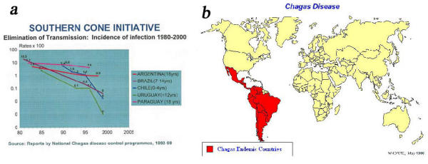
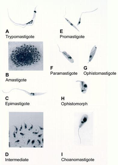
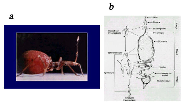
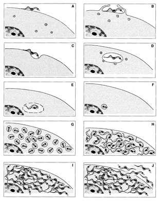
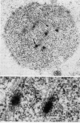
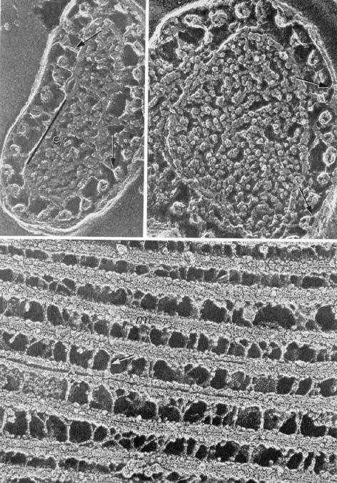
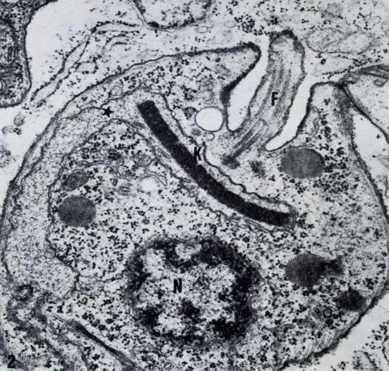
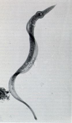
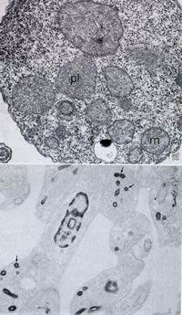
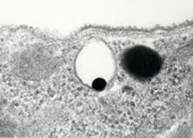
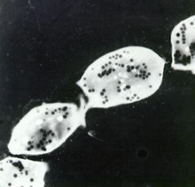
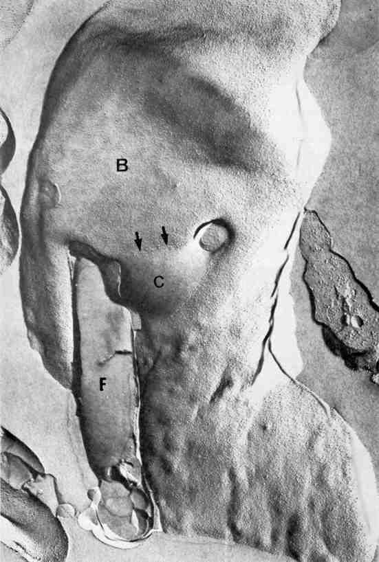
Members of the Trypanosomatidae family comprise a large number of species that are causative agents of important diseases such as sleeping sickness, Chagas' disease and Leishmaniasis. These organisms are also of biological interest since they are able to change the morphology according to the environment where they live, through a process of reversible cell transformation, and possess structures and organelles that are not found in mammalian cells. This review analyses the process of transformation, which takes place during the life cycle of Trypanosoma cruzi in the vertebrate and invertebrate hosts. Special attention is given to the interaction of the parasite with vertebrate cells. In addition, the present knowledge of structures and organelles such as the nucleus, the plasma membrane, the sub-pellicular microtubules, the flagellum, the kinetoplast-mitochondrion complex, the peroxisome (glycosome), the acidocalcisome and the structures and organelles involved in the endocytic pathway, is reviewed from a cell biology perspective. The possible use of available data for the development of new anti parasite drugs is also discussed.


Figures

Figure 1
Figure 1
a: Elimination of transmission of Chagas' disease. b: Distribution of Chaga's disease in the world.
Figure 2
Figure 2
Light microscopy of stained cells showing the developmental stages found in the members of the Trypanosomatidae family.
Figure 3
Figure 3
a: View of a triatomine after blood meal (courtesy of P Azambuja and E Garcia). b: Schematic view of the development of Trypanosma cruzi within the invertebrate host.
Figure 4
Figure 4
Schematic view of the various phases of the interaction of T. cruzi with vertebrate cells.
Figure 5
Figure 5
Two different views of the interaction of T. cruzi with macrophages, as seen by scanning electron microscopy.
Figure 6
Figure 6
Trypomastigote form of T. cruzi a few hours after penetration into the host cell. Rupture of the membrane lining the parasitophorous vacuole can be seen (arrows).
Figure 7
Figure 7
Different views of the membrane lining recem penetrated trypomastigote forms of T. cruzi.
Figure 8
Figure 8
Freeze-fracture view of an epimastigote form of T. cruzi. The nuclear pores are evident.
Figure 9
Figure 9
Low and high magnification of a nucleus of an epimastigote form of T. cruzi in process of division. (courtesy of A Solari).
Figure 10
Figure 10
Different views of the association of the sub-pellicular microtubules with each other and with the plasma membrane.
Figure 11
Figure 11
Amastigote form of T. cusi as seen in a thin section. A short flagellum is evident.
Figure 12
Figure 12
High voltage electron microscopy showing an intact tyrypomastigote form of T. cruzi.
Figure 13
Figure 13
Different views of the attachment of the flagellum to the cell body of T. cruzi as seen in freeze-fracture replicas.
Figure 14
Figure 14
a and b: Two different views of the paraflagellar structure as seen in thin sections and in replicas of quick frozen, freeze-fracture and deep-etched cells.
Figure 15
Figure 15
Deep-etchinng view of the kinetoplast and the region of flagellar emergence of an epimastigote form of T. cruzi.
Figure 16
Figure 16
General view of an epimastigote form of T. cruzi incubated in the presence of a drug which inhibits the biosynthesis of ergosterol. An intense swelling of the mitochondrion is observed.
Figure 17
Figure 17
Two views of the glycosomes in trypanosomatids as seen in a conventional thin section and in cells incubated in the presence of ethanolic phosphotungstic acid, which reveals basic proteins.
Figure 18
Figure 18
Visualization of an acidocalcisome of T. cruzi as seen in a thin section.
Figure 19
Figure 19
Electron spectroscopic image of whole amastigote forms of T. cruzi. The acidocalcisomes appear as electron-dense structures.
Figure 20
Figure 20
General view of an epimastigote form of T. cruzi as seen in a freeze-fracture replica. The flagellar membrane and the cytostome can be seen.
Figure 21
Figure 21
Thin section showing the initial internalization of proteins through the cytostome (arrow).
Figure 22
Figure 22
Reservosomes found in the epimastigote form of T. cruzi.
Figure 23
Figure 23
Immunocytochemical localization of cysteine proteinase in an epimastigote form of T. cruzi. Intense labeling of the cell surface and reservosomes can be observed.

References

    1. Briones MRS, Souto RP, Stolf BS, Zingales B. The evolution of two Trypanosoma cruzi subgroups inferred from rRNA genes can be correlated with the interchange of American mammalian faunas in the Cenozoic and has implications to pathogenicity and host specificity. Mol Biochem Parasitol. 1999;104:219–232. doi: 10.1016/S0166-6851(99)00155-3. - DOI - PubMed
    1. Brisse S, Verhoef J, Tibayrenc M. Characterisation of large and small subunit rRNA and mini-exon genes further supports the distinction of six Trypanosoma cruzi lineages. Int J Parasitol. 2001;31:1218–1226. doi: 10.1016/S0020-7519(01)00238-7. - DOI - PubMed
    1. Carvalho TU, De Souza W. Infectivity of amastigotes of Trypanosoma cruzi. Rev Inst Med Trop São Paulo. 1986;28:205–212. - PubMed
    1. Ley V, Andrews NW, Robbins ES, Nussenszweig V. The exit of Trypanosoma cruzi from the phagosome is inhibited by raising the pH of acidic compartments. J Exp Med. 1990;168:649–659. - PMC - PubMed
    1. Meyer H, De Oliveira M. Cultivation of Trypanosoma cruzi in tissue cultures: a four year study. Parasitology. 1948;39:91–94. - PubMed

LinkOut - more resources