Case Reports
. 2002 Oct;86(10):1093-8.
doi: 10.1136/bjo.86.10.1093.

Clinicopathological correlation of polypoidal choroidal vasculopathy revealed by ultrastructural study

Affiliations
Case Reports

Clinicopathological correlation of polypoidal choroidal vasculopathy revealed by ultrastructural study

A Okubo et al. Br J Ophthalmol. 2002 Oct.

Abstract

Aims: To describe the clinical and histopathological findings in a patient with polypoidal choroidal vasculopathy.

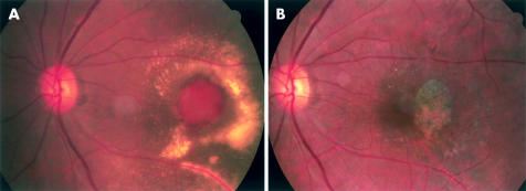
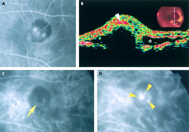
Methods: A 76 year old Japanese man had a discrete, orange-red lesion of 1 disc diameter in the macula, with the fluorescein and indocyanine green angiographic and optical coherence tomographic findings compatible with polypoidal choroidal vasculopathy. He underwent a surgical removal of the macular lesion, followed by light and electron microscopic examinations.

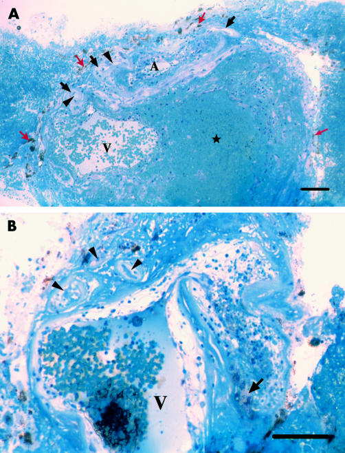
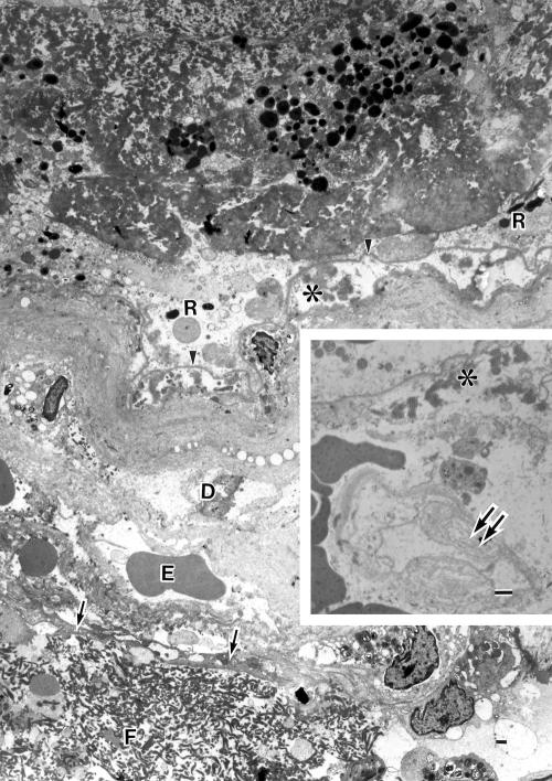
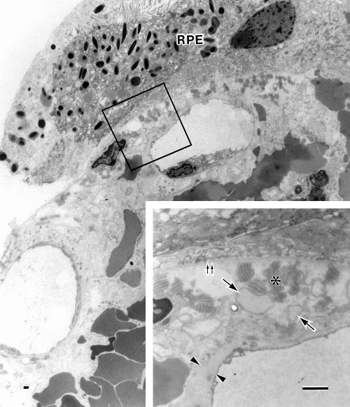
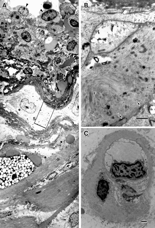
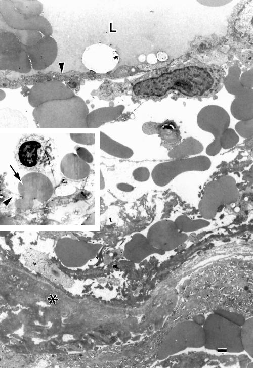
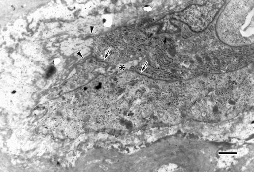
Results: The histopathological examination revealed that the specimen consisted of degenerated retinal pigment epithelium-Bruch's membrane-choriocapillaris complex and inner choroid. A tortuous, unusually dilated venule was present adjacent to an arteriole with marked sclerotic changes, appearing to form arteriovenous crossing. These vessels seemed to represent native inner choroidal vessels, and had haemorrhage per diapedesis. Blood cells and fibrin filled the lumina of the vessels and accumulated in the extravascular spaces, indicating vascular stasis.

Conclusion: Hyperpermeability and haemorrhage due to stasis of a dilated venule and an arteriole involved by sclerosis at the site where they cross in the inner choroid might cause oedema and degeneration of the tissue. Voluminous accumulation of blood cells and fibrin might generate elevation of tissue pressure sufficient to displace the weakened lesion anteriorly. The result suggests that the polypoidal vessels in this case represent abnormality in the inner choroidal vasculature.


Figures

Figure 1
Figure 1
Colour fundus photographs of the left eye. (A) Fundus photograph shows an elevated, oval, 1 disc diameter size, orange-red lesion showing several polypoidal structures in the central portions with an overlying haemorrhagic pigment epithelial detachment and subretinal haemorrhage. There is a ring of exudates surrounding the neurosensory retinal detachment which overlies the lesion. (B) Eight months after operation there is an RPE defect slightly temporal to the foveola.
Figure 2
Figure 2
The left eye of the patient. (A) Fluorescein angiogram taken 58 seconds after dye injection shows several hyperfluorescent spots, some of which are leaking, in the macula surrounded by blocked fluorescence. (B) Optical coherence tomographic image scanning the orange-red lesion demonstrates anterior bulging of highly reflective layers which comprise the sensory retinal layer and the surface layer (arrowhead) of the bulged tissue, shadowing the underlying portion. There is a low reflective space (asterisk) suggestive of serous retinal detachment. (C) Indocyanine green angiogram taken 23 seconds after dye injection demonstrates a faint, horseshoe-shaped fluorescence of a large vessel (arrow) in the lesion. (D) Indocyanine green angiogram after 68 seconds. A tortuous vascular structure with polyp-like structures (arrowheads) is seen near, or overlapping with, the horseshoe-shaped vessel.
Figure 3
Figure 3
Photomicrographs of approximately vertically sectioned separate portions of the excised specimen (toluidine blue staining). (A) Degenerate pigment containing cells (red arrows), hyaline-like layer (black arrows), and capillaries (arrowheads) occupy its inner surface. A large venule (V) with extravasated blood cells in the wall lies adjacent to an arteriole (A). Blood cells and fibrin are accumulated within, and on the lateral surface of the specimen. Bar = 100 μm. (B) A tortuous venule (V) shows bent and dilated portions, whose wall is oedematous with extravasated blood cells and lipid laden cells (arrow). The lumen is filled with blood cells and masses of fibrin. Degenerate small arterioles and capillaries (arrowheads) with thickened basement membrane and obstructed lumen lies adjacent to the venule. Bar = 100 μm.
Figure 4
Figure 4
Electron micrograph showing the hyaline-like layer on the surface of the lesion. The basement membrane (arrowheads) of the pigment containing cells (R) is continuous and unusually electron dense. A few collagen fibrils, many wide spaced collagen (asterisk) and vesicular/linear deposits exist beneath the basement membrane. A portion of the vitreal side wall of a large arteriole is seen which is a serial section of the large arteriole (A) in Figure 3A (a portion of the scleral side wall of this vessel is shown in Fig 6). The arteriole, whose endothelium is indicated by arrows, contains blood cells and fibrin masses (F) in lumen. The seriously degenerated wall consists almost entirely of basement membrane-like substances and remnants of some degenerated cells (D), comprising the outer portions of the hyaline-like layer. Erythrocytes (E) are extravasated in the wall around the endothelium. Inset: A ghost choriocapillary (double arrows) with basement membrane remnant is seen beneath the degenerated Bruch’s membrane containing wide spaced collagen (asterisk). Bar = 1 μm.
Figure 5
Figure 5
Electron micrograph of a peripheral surface portion of the specimen. The hyaline-like layer is present between the retinal pigment epithelial cells (RPE) and the choriocapillaris that show nearly normal structures. Extravasated erythrocytes are seen mainly external to the choriocapillaris and occasionally in the intercapillary zone. Inset: Magnification of the enclosed area. Bruch’s membrane is oedematous and full of wide spaced collagen (asterisk) and vesicular profiles (arrows), showing no normal structures such as elastic layer. Double arrows: Basement membrane of the RPE. The endothelium of the choriocapillaris shows fenestrae and thick basement membrane (arrowheads). Bar = 1 μm.
Figure 6
Figure 6
Electron micrographs of a degenerate arteriole and a capillary. (A) A portion of the scleral side wall of the degenerate large arteriole, showing platelets and erythrocytes seen around its endothelial cells (arrowheads), increased basement membrane substances (asterisks), variably degenerate smooth muscle cells (S), and lipid laden cell (L). (B) Magnification of the enclosed area in (A), showing smooth muscle cells with myofilaments (F) and sublemmal dense bodies (arrows). (C) A choriocapillaris with decreased fenestrae and thickened basement membrane. Bar = 1 μm.
Figure 7
Figure 7
Electron micrograph of a portion of the degenerate wall of a dilated large venule. The wall is oedematous with extravasated erythrocytes and fibrin around endothelium (arrowhead). L = lumen; asterisk = basement membrane substances. Inset: an erythrocyte (arrow) is passing through a gap between endothelial cells (arrowhead). Bar = 1 μm.
Figure 8
Figure 8
Electron micrograph of a newly formed vessel located near the large dilated venule. The plump endothelial cells with discontinuous thin basement membranes (arrowheads) are bound each other by immature intercellular junction (arrows), and have formed immature lumen (asterisk). Bar = 1 μm.

References

    1. Stern RM, Zakov ZN, Zegarra H, et al. Multiple recurrent serosanguineous retinal pigment epithelial detachments in black woman. Am J Ophthalmol 1985;100:560–9. - PubMed
    1. Yannuzzi LA, Sorenson J, Spaide RF, et al. Idiopathic choroidal vasculopathy (IPCV). Retina 1990;10:1–8. - PubMed
    1. Kleiner RC, Brucker AJ, Johnston RL. The posterior uveal bleeding syndrome. Retina 1990;10:9–17. - PubMed
    1. Perkovich BT, Zakov ZN, Berlin LA, et al. An update on multiple recurrent serosanguineous retinal pigment epithelial detachments in black woman. Retina 1990;10:18–26. - PubMed
    1. Spaide RF, Yannuzzi LA, Slakter JS, et al. Indocyanine green videoangiography of idiopathic polypoidal choroidal vasculopathy. Retina 1995;15:100–10. - PubMed

Publication types