

Measurement of optic nerve sheath diameter by ultrasound: a means of detecting acute raised intracranial pressure in hydrocephalus
- PMID: 12234888
- PMCID: PMC1771326
- DOI: 10.1136/bjo.86.10.1109
Measurement of optic nerve sheath diameter by ultrasound: a means of detecting acute raised intracranial pressure in hydrocephalus
Abstract
Aim: To evaluate the utility of measuring the optic nerve sheath diameter in children with shunted hydrocephalus, suspected of having raised intracranial pressure.
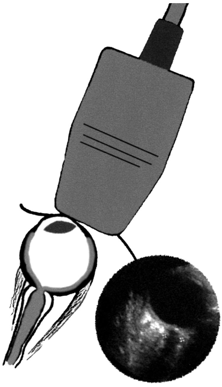
Methods: 23 children with shunted hydrocephalus were examined, six had well controlled ICP, 17 however manifested symptoms suggestive of intracranial hypertension. A clinical history was taken from all patients and their parents or carers. The shunt valve was examined clinically, and signs of raised intracranial pressure were sought. Ultrasound examination was performed in both eyes to measure the optic nerve sheath diameters 3 mm behind the globe. These measurements were compared with control data obtained from 102 children who attended the radiology department for unrelated renal ultrasound examination.
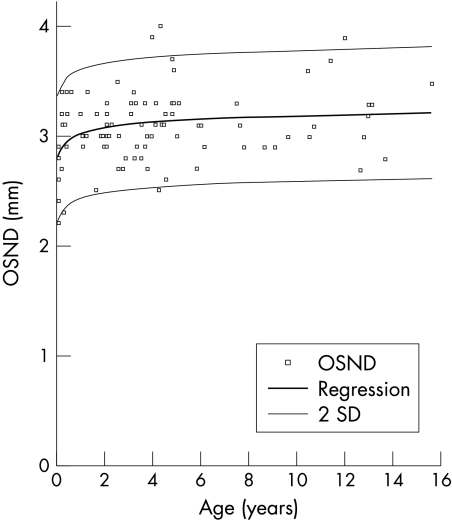
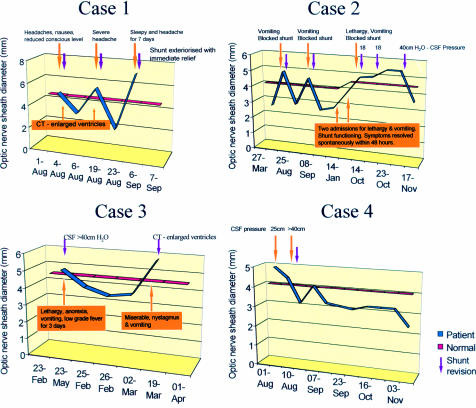
Results: Control data suggested that the upper limit of normal for optic nerve sheath diameter is 4.5 mm (measured 3 mm behind the globe) in patients over 1 year of age, and 4.0 mm in children less than 1 year of age. Those patients with functioning ventriculoperitoneal shunts had a mean optic nerve sheath diameter of 2.9 (SD 0.5) mm; those with raised intracranial pressure had a mean optic nerve sheath diameter of 5.6 (0.6) mm (p<0.0001). These results confirm that optic nerve sheath diameters in excess of the control data are strongly suggestive of raised intracranial pressure.
Conclusion: The evaluation of the optic nerve sheath diameter is a simple non-invasive procedure, which is a potentially useful tool in the assessment and monitoring of children with hydrocephalus suspected of having raised intracranial pressure.
Figures
References
-
- Helmke H, Hansen HC. Fundamentals of transorbital sonographic evaluation of optic nerve sheath expansion under intracranial hypertension. I Experimental study. Paediatr Radiol 1996;26:701–5. - PubMed
-
- Helmke H, Hansen HC. Fundamentals of transorbital sonographic evaluation of optic nerve sheath expansion under intracranial hypertension. II Patient study. Paedaitr Radiol 1996;26:706–10. - PubMed
-
- Ballantyne J, Hollman AS, Hamilton R, et al. Transorbital optic nerve sheath ultrasonography in normal children. Clin Radiol 1999;54:740–2. - PubMed
-
- Liu D, Kahn M. Measurement and relationship of subarachnoid pressure of the optic nerve to intracranial pressure in fresh cadavers. Am J Ophthalmol 1993;116:548–56. - PubMed
-
- Ossoinig KC. Standardised echography: basic principles, clinical applications and results. Int Ophthalmol Clin 1979;19:127–210. - PubMed
MeSH terms
LinkOut - more resources
Full Text Sources
Other Literature Sources
Medical