. 2002 Sep 16;21(18):4796-808.
doi: 10.1093/emboj/cdf493.

Sprouty2 attenuates epidermal growth factor receptor ubiquitylation and endocytosis, and consequently enhances Ras/ERK signalling

Affiliations

Sprouty2 attenuates epidermal growth factor receptor ubiquitylation and endocytosis, and consequently enhances Ras/ERK signalling

Esther Sook Miin Wong et al. EMBO J. .

Abstract

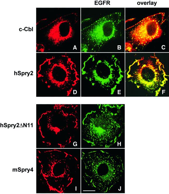
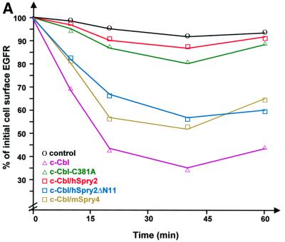
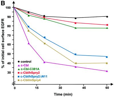
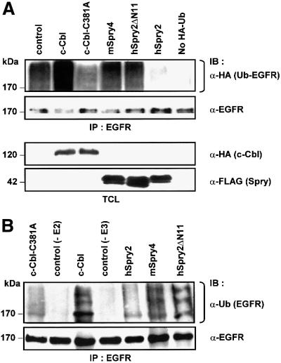
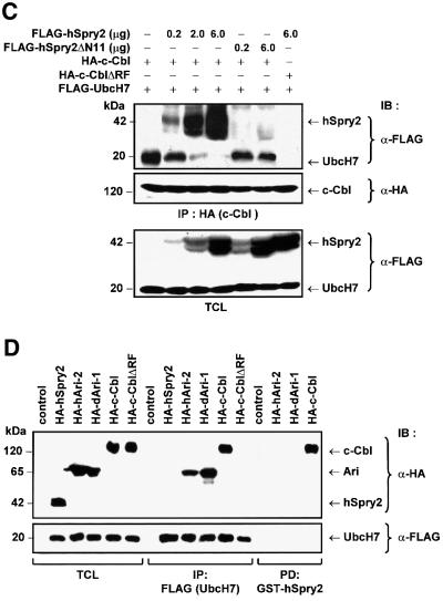
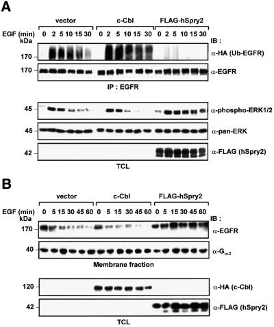
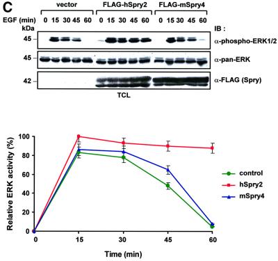
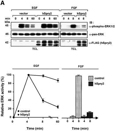
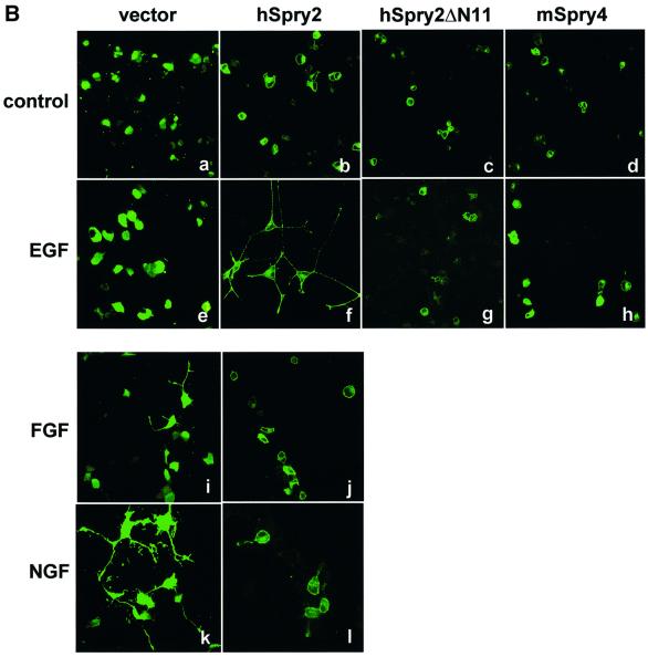
Drosophila Sprouty (dSpry) was genetically identified as a novel antagonist of fibroblast growth factor receptor (FGFR), epidermal growth factor receptor (EGFR) and Sevenless signalling, ostensibly by eliciting its response on the Ras/MAPK pathway. Four mammalian sprouty genes have been cloned, which appear to play an inhibitory role mainly in FGF- mediated lung and limb morphogenesis. Evidence is presented herein that describes the functional implications of the direct association between human Sprouty2 (hSpry2) and c-Cbl, and its impact on the cellular localization and signalling capacity of EGFR. Contrary to the consensus view that Spry2 is a general inhibitor of receptor tyrosine kinase signalling, hSpry2 was shown to abrogate EGFR ubiquitylation and endocytosis, and sustain EGF-induced ERK signalling that culminates in differentiation of PC12 cells. Correlative evidence showed the failure of hSpry2DeltaN11 and mSpry4, both deficient in c-Cbl binding, to instigate these effects. hSpry2 interacts specifically with the c-Cbl RING finger domain and displaces UbcH7 from its binding site on the E3 ligase. We conclude that hSpry2 potentiates EGFR signalling by specifically intercepting c-Cbl-mediated effects on receptor down-regulation.


Figures

None
Fig. 1. hSpry2 specifically inhibits EGFR endocytosis. COS-1 cells were singly transfected with 1 µg each of HA-tagged c-Cbl alone or FLAG-tagged Sprys, and subjected to serum-deprivation overnight prior to stimulation with 100 ng/ml EGF at 37°C for 10 min. Cells were then fixed, permeabilized and stained for endogenous EGFR using an anti-EGFR monoclonal and FITC-conjugated AffiniPure rabbit anti-mouse IgG (green) secondary antibody. c-Cbl was visualized with an anti-c-Cbl polyclonal, and FLAG-tagged Spry constructs were detected using a polyclonal anti-FLAG, followed by Texas Red dye-conjugated AffiniPure goat anti-rabbit IgG (red). Merged signals are labelled as overlay staining (yellow). Bar = 10 µm.
None
Fig. 2. hSpry2 enhances cell surface retention of EGFRs. (A) To quantify the surface EGFR population, COS-1 cells were co-transfected with 0.1 µg of an EGFR expression vector, together with 0.5 µg plasmid encoding c-Cbl alone (pink triangles) or with either 0.4 µg FLAG–hSpry2 (red squares), FLAG–hSpry2ΔN11 (blue squares) or FLAG–mSpry4 (yellow squares) cDNA, or with 1.0 µg plasmid encoding the RING finger-defective form of c-Cbl (C381A) alone (green triangles). Forty-eight hours post-transfection, serum-starved culture cells were subjected to stimulation without or with 100 ng/ml EGF at 37°C for the time periods indicated. Bound EGF was removed, and the level of surface receptors relative to the initial number of ligand-binding sites was determined by incubating sister cultures with [125I]EGF (10 ng/ml) at 4°C for 2–4 h, in the absence or presence of a 100-fold excess of unlabelled EGF (1 µg/ml). Control cells were not exposed to EGF (circles). The results are expressed as the average fraction of original binding sites that remained on the cell surface after exposure to the unlabelled ligand at 37°C. The graphical plot shown is derived from a single experiment that is representative of four independent experiments. (B) Similar transfections (closed symbols) and treatment as in (A), except that cells were pre-incubated without or with 100 ng/ml EGF in the presence of 30 µM monensin at 4°C for 2 h before a temperature shift to 37°C for the various time periods. The residual level of surface-bound receptors that did not undergo down-regulation was then assayed by performing a direct binding assay with radiolabelled EGF. The average of duplicate determinations was expressed as the percentage of total radioactivity at time = 0 min. The experiment was repeated twice.
None
Fig. 3. hSpry2 abrogates c-Cbl-dependent ubiquitylation of EGFRs. (A) COS-1 cells were co-transfected with 1.0 µg HA-tagged ubiquitin and 3.0 µg each of either vector control, c-Cbl, c-Cbl-C381A or the different FLAG-tagged Spry forms per 100 mm culture dish. Forty-eight hours later, cell monolayers were treated with 100 ng/ml EGF at 37°C for 10 min. Total cell lysates (TCL) were subjected to immunoprecipitation (IP) using anti-EGFR–agarose-conjugated beads, immunoblotted (IB) with anti-HA to distinguish ubiquitin-conjugated EGFR, and anti-EGFR to assess the amounts of immunoprecipitated EGFR. TCL samples were analysed with anti-HA or anti-FLAG to show relative expression levels of the various constructs. The bracket indicates the position of high molecular weight species of ubiquitin-positive EGFRs. (B) Immunoprecipitated EGFR proteins were subjected to an in vitro ubiquitylation assay in the presence of purified UbcH7 (or no added UbcH7 as control without E2), eluted c-Cbl protein alone (or no added c-Cbl as control without E3), recombinant c-Cbl with either GST–hSpry2, GST–mSpry4 or GST–hSpry2ΔN11 fusion proteins, or c-Cbl-C381A fusion protein alone, plus the essential components in the ubiquitylation system. The reaction products were analysed following western blotting protocol with anti-ubiquitin. The bracket highlights the position of high molecular species of ubiquitin-positive EGFRs. (C) Competitive binding between hSpry2 and UbcH7 for the RING finger domain of c-Cbl. COS-1 cells (100 mm dishes) were co-transfected with 3.0 µg each of FLAG-tagged UbcH7, HA–c-Cbl or HA–c-Cbl-ΔRF mutant, and varying amounts of FLAG–hSpry2 or the c-Cbl non-binding truncation mutant FLAG–hSpry2ΔN11, as indicated. Serum-deprived cells were stimulated with 100 ng/ml EGF at 37°C for 10 min. TCL were subjected to precipitation using anti-HA and immunoblotted (IB) with anti-FLAG to detect associated hSpry2 or UbcH7, and anti-HA to show the relative amounts of immunoprecipitated c-Cbl. A TCL blot was probed with anti-FLAG to demonstrate quantitatively the expression levels of exogenous hSpry2 and UbcH7 proteins. (D) COS-1 cells (100 mm dishes) were untransfected (control), or co-transfected with 3.0 µg each of FLAG-tagged UbcH7 and either HA–hSpry2, HA–human Ariadne-2 (hARI-2), HA–Drosophila Ariadne-1 (dAri-1), HA–c-Cbl or HA–c-CblΔRF constructs. Immunocomplexes of FLAG–UbcH7 or ‘pull-downs’ using GST–hSpry2 were detected using anti-HA. Total cell lysates were analysed for equivalent levels of protein expression and normalized sample loading using antibodies as indicated. An immunoblot (with TCL loaded alongside) was probed with anti-HA to detect hSpry2-binding proteins, and with anti-FLAG to verify equal amounts of immunoprecipitated UbcH7.
None
Fig. 4. hSpry2 elicits sustained ERK activation in response to EGF induction. (A) 293T cells (60 mm dishes) were co-transfected with plasmids encoding EGFR (0.2 µg) and HA-tagged ubiquitin (0.2 µg), together with an empty vector control, HA–c-Cbl (1.0 µg) or FLAG–hSpry2 (0.8 µg). Forty-eight hours post-transfection, cell monolayers were incubated with 100 ng/ml EGF at 37°C for the indicated time periods. Total cell lysates (TCL) were subjected to precipitation using anti-EGFR–agarose-conjugated beads, and receptor ubiquitylation was detected with an anti-HA probe against HA–Ub-conjugated EGFR. An anti-EGFR blot demonstrates equal amounts of immunoprecipitated EGFR. Total cell extracts were immunoblotted (IB) with anti-phospho-ERK1/2 (p44/42) to detect corresponding activated endogenous ERK levels; anti-pan-ERK to ascertain equivalent protein loading; and anti-FLAG to assess equality of expression of FLAG–hSpry2. (B) Cell membrane extracts of a similar set of transfections to that depicted in (A) were isolated and immunoblots of one-tenth sample volumes loaded were assayed with anti-EGFR to assess the levels of surface EGFRs; and with anti-Giα3 to ascertain equivalent amounts of loaded membrane fraction samples. TCL were analysed for protein expression of transfected c-Cbl and hSpry2 by immunoblotting (IB) with anti-HA and anti-FLAG, respectively. (C) 293T cells (60 mm dishes) co-transfected with 0.2 µg EGFR and 0.8 µg of either vector control, FLAG–hSpry2 or FLAG–mSpry4 were treated with 100 ng/ml EGF at 37°C for the different time periods, as indicated. TCL were immunoblotted (IB) with anti-phospho-ERK1/2 to assess the activation profiles of endogenous ERK. Anti-pan-ERK and anti-FLAG were used to reveal the equality of protein loading and expression. Phospho-ERK1/2 signals were quantified with the aid of a densitometer, and represented as a percentage of maximal ERK activity (ERK activation, for hSpry2, at 15 min stimulation treated as 100%) in the graphical plot. Shown are the averages of three separate experiments ± SEM.
None
Fig. 5. (A) hSpry2 potentiates EGF- but inhibits FGF-induced ERK activation. PC12 cells (60 mm dishes) were transiently transfected with 2.0 µg each of FLAG vector or FLAG–hSpry2. Forty-eight hours post-transfection, cells were adjusted to low serum condition (1.0% HBS, 0.5% FBS) for 3–5 h before stimulation with 100 ng/ml EGF or 10 ng/ml FGF at 37°C for the time periods indicated. TCL were resolved by immunoblotting (IB) analysis to detect activated ERK (p42/44) using anti-phospho-ERK1/2, and probed with anti-pan-ERK and anti-FLAG to show similar protein loading and expression of the transfected Spry proteins. The amount of activated ERK1/2 signals was measured on a PhosphorImager, and presented as a percentage of maximal ERK activity at 4 min (EGF) and 8 min (FGF) stimulation in the respective line or bar chart. Shown are the averages of three independent experiments ± SEM. (B) EGF-induced neurite extensions are observed in PC12 cells expressing hSpry2. PC12 cells transfected with 2.0 µg plasmids encoding either GFP–vector control, GFP–hSpry2, GFP–hSpry2ΔN11 or GFP–mSpry4 were grown on poly-lysine-coated cover slips, and fixed after 4 days incubation in low serum media without added growth factor (control), supplemented with 100 ng/ml EGF, 10 ng/ml FGF or 10 ng/ml NGF. Panels show GFP fluorescence (green) of representative cell clones from each experimental treatment.

Similar articles

Cited by

References

    1. Ardley H.C., Tan,N.G., Rose,S.A., Markham,A.F. and Robinson,P.A. (2001) Features of the Parkin/Ariadne ubiquitin ligase, HHARI, which regulates its interaction with the ubiquitin-conjugating enzyme, UbcH7. J. Biol. Chem., 276, 19640–19647. - PubMed
    1. Aguilera M., Oliveros,M., Martinez-Padron,M., Barbas,J.A. and Ferrus,A. (2000) Ariadne-1: a vital Drosophila gene is required in development and defines a new conserved family of RING Finger proteins. Genetics, 155, 1231–1244. - PMC - PubMed
    1. Bao J., Alroy,I., Waterman,H., Schejter,E.D., Brodie,C., Gruenberg,J. and Yarden,Y. (2000) Threonine phosphorylation diverts internalized epidermal growth factor receptors from a degradative pathway to the recycling endosome. J. Biol. Chem., 275, 26178–26186. - PubMed
    1. Bartkiewicz M., Houghton,A. and Baron,R. (1999) Leucine zipper-mediated homo-dimerization of the adaptor protein c-Cbl. A role in c-Cbl’s tyrosine phosphorylation and its association with epidermal growth factor receptor. J. Biol. Chem., 274, 30887–30895. - PubMed
    1. Basu S.K., Goldstein,J.L., Anderson,R.G. and Brown,M.S. (1981) Monensin interrupts the recycling of low density lipoprotein receptors in human fibroblasts. Cell, 24, 493–502. - PubMed

Publication types

MeSH terms