. 2002 Sep 16;21(18):4863-74.
doi: 10.1093/emboj/cdf495.

Fission yeast Mor2/Cps12, a protein similar to Drosophila Furry, is essential for cell morphogenesis and its mutation induces Wee1-dependent G(2) delay

Affiliations

Fission yeast Mor2/Cps12, a protein similar to Drosophila Furry, is essential for cell morphogenesis and its mutation induces Wee1-dependent G(2) delay

Dai Hirata et al. EMBO J. .

Abstract

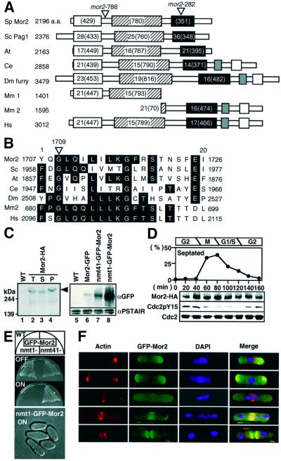
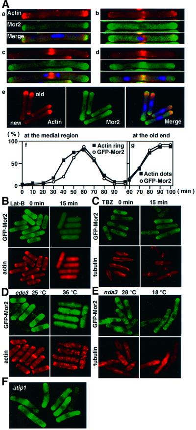
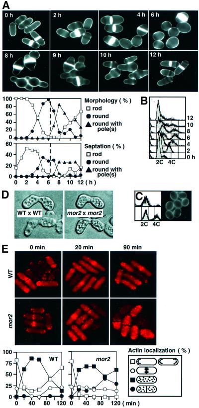
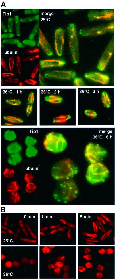
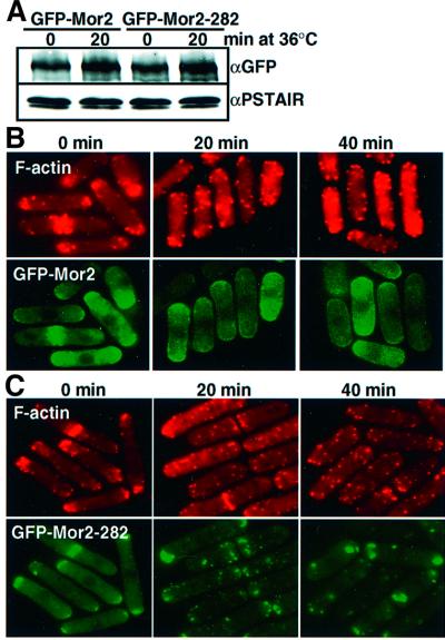
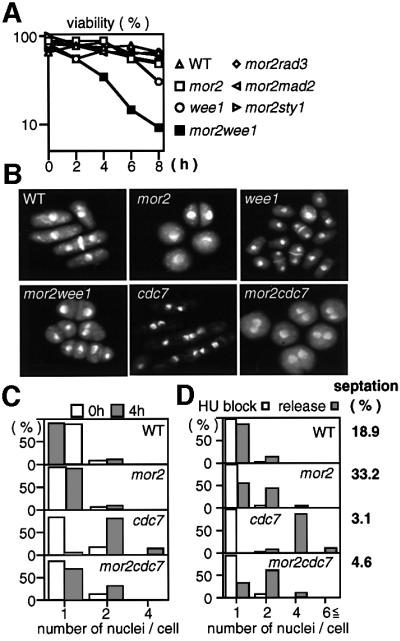
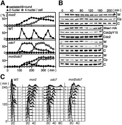
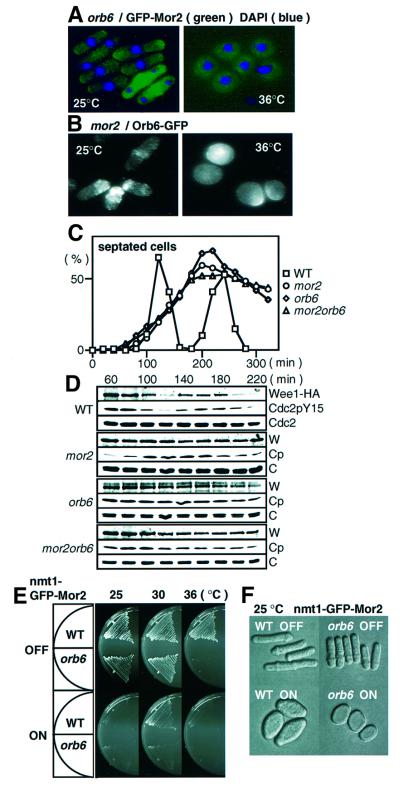
Fission yeast cells identify growing regions at the opposite ends of the cell, producing the rod-like shape. The positioning of the growth zone(s) and the polarized growth require CLIP170-like protein Tip1 and the Ndr kinase Orb6, respectively. Here, we show that the mor2/cps12 mutation disrupts the localization of F-actin at the cell ends, producing spherical cells and concomitantly inducing a G(2) delay at 36 degrees C. Mor2 is important for the localization of F-actin at the cell end(s) but not at the medial region, and is essential for the restriction of the growth zone(s) where Tip1 targets. Mor2 is homologous to the Drosophila Furry protein, which is required to maintain the integrity of cellular extensions, and is localized at both cell ends and the medial region of the cell in an actin-dependent fashion. Cellular localization of Mor2 and Orb6 was interdependent. The tyrosine kinase Wee1 is necessary for the G(2) delay and maintenance of viability of the mor2 mutant. These results indicate that Mor2 plays an essential role in cell morphogenesis in concert with Orb6, and the mutation activates the mechanism coordinating morphogenesis with cell cycle progression.


Figures

None
Fig. 1. Identification of Mor2. (A) A schematic presentation of the homology between Mor2 and Furry homologous proteins. Schizosaccharomyces pombe Mor2 is aligned with S.cerevisiae Pag1/Tao3 (Sc), Arabidopsis thaliana T51397 (At), C.elegans AAF99910 (Ce), D.melanogaster Furry (Dm), Mus musculus CAC42175.1 (Mm1), CAC42196.1 (Mm2) and Homo sapiens CAB42442 (Hs). Identities (%) and the number of amino acids are indicated. (B) The mutation site of the mor2-282 allele. (C) Identification of Mor2 protein. Lanes 1–4: total cell extract prepared from wild type (WT, 972), and the cells having mor2+:3HA:kanr (Mor2-HA, YS78-1) grown in YPD medium were electrophoresed on SDS–PAGE gels and immunoblotted with anti-HA (lanes 1 and 2). Total cell extracts (T, lane 2) prepared from the Mor2-HA strain were fractionated into soluble (S, lane 3) and insoluble fractions (P, lane 4) by centrifugation at 14 000 g. Lanes 5–8: wild type, and cells with mor2+:GFP:kanr (Mor2–GFP, YS77), kanr:nmt41-GFP:mor2+ (nmt41–GFP–Mor2, YS84-1) or kanr:nmt1-GFP:mor2+ (nmt1-GFP–Mor2, YS85) grown in Edinburgh minimal medium (EMM) containing 4 µM thiamine were transferred into EMM. After cultivation for 14 h, the cells were collected. Total cell extracts prepared from the cells were immunoblotted with anti-GFP or anti-PSTAIR antibodies. (D) Mor2 protein during the cell cycle. Early G2 cells of Mor2-HA strain were collected by centrifugal elutriation and cultured in YPD medium at 28°C. Samples of the culture were taken at the indicated times for calculating the percentage of septated cells. Total cell extracts prepared from the samples of the same cultures were immunoblotted with anti-HA (for Mor2-HA), anti-PSTAIR (for Cdc2) or anti-Cdc2 phosphorylated on tyrosine-15 (for Cdc2-P) antibodies. (E) Growth (upper) and morphology (lower) of the Mor2-over-expressing cells. Upper panel: the nmt1-GFP–Mor2 and nmt41–GFP–Mor2 strains were cultured on EMM (ON) or EMM plates containing 4 mM thiamine (OFF) at 25°C for 3 days. Lower panel: the nmt1-GFP–Mor2 strain grown in EMM containing 4 µM thiamine was transferred into EMM medium and cultured for 18 h at 25°C. (F) Localization of Mor2 and F-actin. The nmt41–GFP–Mor2 strain cells grown in EMM containing 4 µM thiamine were transferred into EMM medium. After cultivation for 14 h, cells were fixed and stained with rhodamine– phalloidin. Merged image (Merge): F-actin (red), GFP–Mor2 (green) and DAPI (blue).
None
Fig. 2. Role of cytoskeletons in Mor2 localization. (A) Mor2 localization in the cells released from G2 block. The cdc25-22 mutant cells having kanr:nmt41-GFP:mor2+ (DH294-4A) grown in EMM containing 8 µM thiamine were transferred into EMM medium. After cultivation for 11–12 h at 25°C, the cells were transferred to 36°C and kept there for 4 h. The medium was then was returned to 25°C, and the cells were fixed and stained with rhodamine–phalloidin (a–e). The appearance of the actin ring at the medial region (f) and actin dots at the old end (g) was examined with respect to Mor2 accumulation at their sites in the time course. (B and C) Effect of Lat-B and TBZ on Mor2 localization. The nmt41–GFP–Mor2 strain (YS84-1) cells grown in EMM containing 8 µM thiamine were transferred into EMM medium. After culti vation for 14–15 h at 25°C, the cells were incubated with 100 µM Lat-B (B) or 100 µM TBZ (C). Cells taken at the indicated times were fixed and stained with rhodamine–phalloidin (B) or anti-tubulin antibody (C). (D) Mor2 localization in the cdc3 mutant. The cdc3-6 (profilin) mutant cells with kanr:nmt41-GFP:mor2+ (DH299-2) grown in EMM containing 8 µM thiamine were transferred into EMM medium. After cultivation for 12 h at 25°C, the cells were transferred to 36°C for 3–4 h, fixed, and stained with rhodamine–phalloidin. (E) Mor2 localization in the nda3 mutant. The nda3-KM311 (β-tubulin) mutant cells with kanr:nmt41-GFP:mor2+ (DH297-1C) grown in EMM containing 8 µM thiamine were transferred into EMM medium. After cultivation for 11–12 h at 28°C, the cells were transferred to 18°C for 6 h, fixed, and immunostained with anti-tubulin antibodies. (F) Mor2 localization in tip1-deleted cells. The Δtip1 mutant cells with kanr:nmt41-GFP:mor2+ (DH421-2B) grown in EMM containing 8 µM thiamine were transferred into EMM medium and cultivated for 13 h at 25°C.
None
Fig. 3. Phenotypes of the mor2 mutant. (A and B) Reversible phenotype of the mor2 mutant. mor2 mutant cells (DH107-4C) grown in YPD medium at 25°C were transferred to 36°C (time 0 h). After incubation for 6 h, the cells were shifted down to 28°C and incubated for 6 h (total 12 h). The cells at the indicated times were taken for observation of cell morphology after having been stained with Calcofluor (A) and for FACS analysis (B). (Cmor2 mutant cells (DH107-4C) grown in YPD medium at 25°C were transferred to 36°C and incubated for 4 h. The unseptated round cells in the culture were collected by centrifugal elutriation for FACS analysis (upper panels, the collected round cells; lower panels, the culture) and for observation of morphology (isolated round cells). (D) Shmoo morphology of the mor2 mutant. Wild-type (h) or mor2 mutant cells (h mor2-786) were crossed with wild-type (h+) or mor2 mutant cells (h+ mor2-786), respectively. After incubation for 24 h on malt extract (MEA) plate, the shmoo morphology of the cells was observed. (E) F-actin localization in the mor2 mutant. The wild-type (972) and mor2 mutant cells (DH192-1A) grown in YPD medium at 25°C were transferred to 36°C (time 0 h). The cells were taken at the indicated times for observation of F-actin localization after being stained with rhodamine–phalloidin.
None
Fig. 4. Role of Mor2 in the organization of the MT cytoskeleton. (A) MT structure and Tip1 localization in the mor2 mutant. The mor2 mutant cells (DH107-4C) grown in YPD medium at 25°C were transferred to 36°C. The cells were fixed and immunostained with anti-tubulin and anti-Tip1 antibodies at the times indicated. (B) MT nucleation in the mor2 mutant. Upper panels: the mor2 mutant cells having tip1+:GFP:kanr (DH360-2B) grown in YPD medium at 25°C were incubated on ice for 30 min, re-warmed to 25°C, and collected at the indicated times for immunostaining with anti-tubulin antibodies. Lower panels: the mor2 mutant cells grown in YPD medium at 25°C were transferred to 36°C and kept there for 6 h, incubated on ice for 30 min, and then re-warmed to 36°C and collected at the indicated times.
None
Fig. 5. Localization of the Mor2 mutant protein. (AC) The nmt41–GFP–Mor2 [YS84-1 (B)] and nmt41–GFP–Mor2-282 [YY5-5 (C)] strains grown in EMM containing 8 µM thiamine were transferred into EMM medium. After cultivation for 14 h at 25°C, the cells were transferred to YPD medium at 36°C, fixed at the indicated times, and stained with rhodamine–phalloidin. The cells of the 36°C culture were taken at the indicated times for examination of the GFP–Mor2 and GFP–Mor2-282 proteins by immunoblotting (A).
None
Fig. 6. Wee1-dependent G2 delay in the mor2 mutant. (AC) Viability and nucleus number of the mutant. Wild-type (22), mor2 (DH107-4C), Δwee1 (IY1078), mor2Δwee1 (DH203-6A), mor2Δrad3 (DH205-5D), mor2Δmad2 (DH207-5A), mor2Δsty1 (DH214-3B), cdc7 and mor2cdc7 (DH295-2B) grown in YPD medium at 25°C were transferred to 36°C and collected at the indicated times. The cells were spread on YPD plates, incubated for 3–4 days at 28°C, and assessed for cell viability (A). The nuclear structures of the cells incubated at 36°C for 4 h were observed by DAPI staining (B), and the number of nuclei in the cells at the indicated times was examined (C). (D) Wild-type, mor2, cdc7 and mor2cdc7 mutant cells grown in YPD medium at 25°C were treated with 12 mM hydroxyurea (HU) for 3.5 h and shifted to 36°C for 1 h. HU-treated cells were washed with pre-warmed YPD medium, inoculated into fresh YPD medium, incubated at 36°C for a further 4 h, fixed, and stained with DAPI. HU-treated cells prior to release from HU are labelled ‘HU block’, and cells released from the HU block are labelled ‘release’. The septation index in ‘release’ is indicated.
None
Fig. 7. Maintenance of Wee1 protein levels in the mor2 mutant. (A–C) Early G2 cells of wild type (RA1224), mor2 (DH274-10B), cdc7 (DH306-8A) and mor2cdc7 (DH306-12B) were collected by centrifugal elutriation. The cells were released into YPD medium at 36°C, and taken at the indicated times for observation of cell morphology (A), immunoblotting (B) and FACS analysis (C). The total proteins (100 µg) were electrophoresed on SDS–PAGE gels. Wee1-HA protein, tyrosine phosphorylation of Cdc2, and Cdc2 protein in the samples were analysed by immunoblotting with antibodies specific for the HA epitope (Wee1-HA, W), Cdc2 phosphorylated on tyrosine-15 (Cdc2pY15, Cp) and an anti-PSTAIR antibody (Cdc2, C), respectively.
None
Fig. 8. Functional interaction between Mor2 and Orb6. (A) Localization of Mor2 in the orb6 mutant. The orb6 mutant cells with kanr:nmt41-GFP:mor2+ (DH451-3D) grown in EMM containing 8 µM thiamine were transferred into EMM medium. After cultivation for 12 h at 25°C, the cells were transferred to 36°C and kept there for 6 h. (B) Localization of Orb6 in the mor2 mutant. The mor2 mutant cells with orb6+:GFP:kanr (DH454–2B) and grown in YES5 medium at 25°C were transferred to 36°C and kept there for 6 h; they were then fixed and immunostained with anti-GFP antibodies. (C and D) Early G2 cells of wild type (RA1224), mor2 (DH274-10B), orb6 (DH463-2C) and mor2orb6 (DH436-12B) were collected by centrifugal elutriation. The cells were released into YPD medium at 36°C, and taken at the indicated times to determine frequency of the septated cells (C) and for immunoblotting (D). (E) Overexpression of Mor2 in the orb6 mutant. Wild-type and orb6 mutant cells with kanr:nmt1-GFP:mor2+ (WT, YS85; orb6, DH475-1C) were cultured on EMM (ON) or EMM plates containing 4 µM thiamine (OFF) at the indicated temperatures for 3 days. (F) The strains grown in EMM containing 4 µM thiamine were transferred into EMM medium and cultured for 18 h at 25°C.

References

    1. Alfa C., Fantes,P., Hyams,J., McLeod,M. and Warbrick,E. (1993) Experiments with Fission Yeast: a Laboratory Course Manual. Cold Spring Harbor Laboratory Press, Cold Spring Harbor, NY.
    1. Aligue R., Wu,L. and Russell,P. (1997) Regulation of Schizosaccharomyces pombe Wee1 tyrosine kinase. J. Biol. Chem., 272, 13320–13325. - PubMed
    1. Bahler J., Wu,J., Longtine,M.S., Shah,N.G., McKenzie,A.III, Steever,A.B., Wach,A., Philippsen,P. and Pringle,J.R. (1998) Heterologous modules for efficient and versatile PCR-based gene targeting in Schizosaccharomyces pombe. Yeast, 14, 943–951. - PubMed
    1. Balasubramanian M.K., Hirani,B.R., Burke,J.D. and Gould,K.L. (1994) The Shizosaccharomyces pombe cdc3+ gene encodes a profilin essential for cytokinesis. J. Cell Biol., 125, 1289–1301. - PMC - PubMed
    1. Beinhauer J.D., Hagan,I.M., Hegemann,J.H. and Fleig,U. (1997) Mal3, the fission yeast homologue of the human APC-interacting protein EB-1 is required for microtubule integrity and the maintenance of cell form. J. Cell Biol., 139, 717–728. - PMC - PubMed

Publication types

MeSH terms