. 2002 Sep 16;21(18):4875-84.
doi: 10.1093/emboj/cdf491.

Dual mode of degradation of Cdc25 A phosphatase

Affiliations

Dual mode of degradation of Cdc25 A phosphatase

Maddalena Donzelli et al. EMBO J. .

Abstract

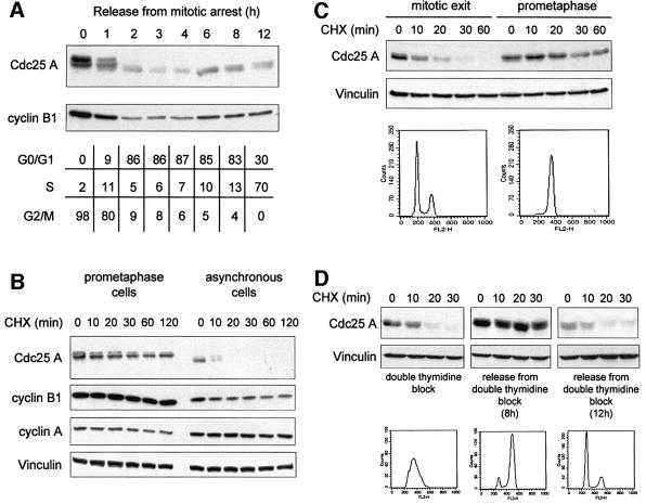
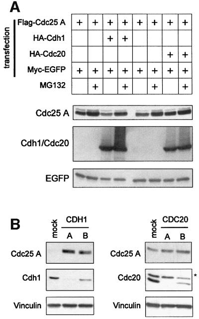
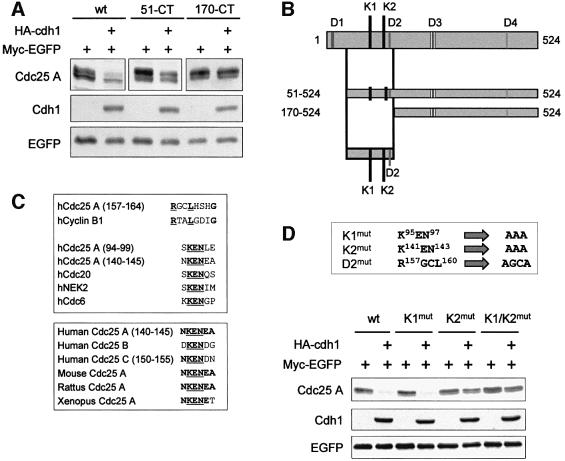
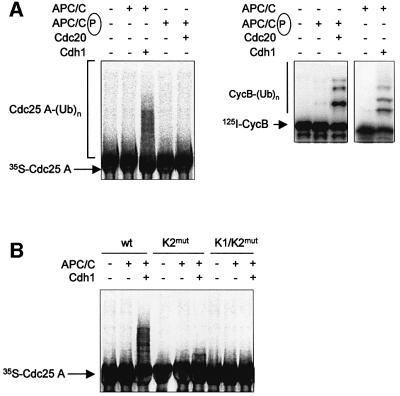
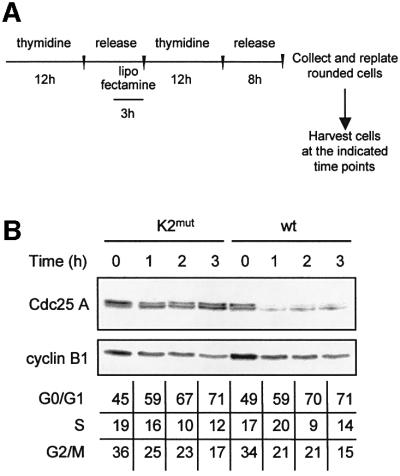
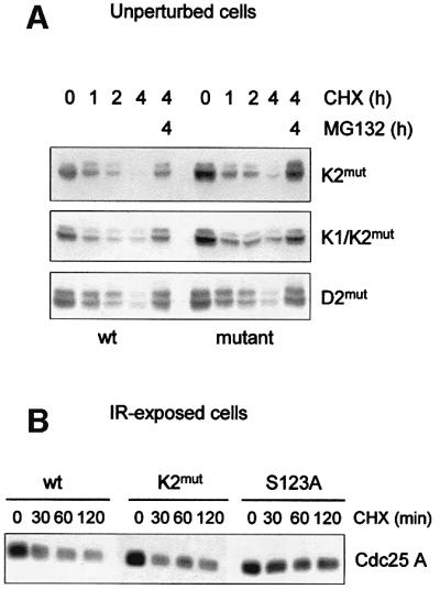
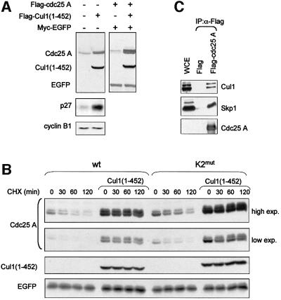
The Cdc25 dual-specificity phosphatases control progression through the eukaryotic cell division cycle by activating cyclin-dependent kinases. Cdc25 A regulates entry into S-phase by dephosphorylating Cdk2, it cooperates with activated oncogenes in inducing transformation and is overexpressed in several human tumors. DNA damage or DNA replication blocks induce phosphorylation of Cdc25 A and its subsequent degradation via the ubiquitin-proteasome pathway. Here we have investigated the regulation of Cdc25 A in the cell cycle. We found that Cdc25 A degradation during mitotic exit and in early G(1) is mediated by the anaphase-promoting complex or cyclosome (APC/C)(Cdh1) ligase, and that a KEN-box motif in the N-terminus of the protein is required for its targeted degradation. Interestingly, the KEN-box mutated protein remains unstable in interphase and upon ionizing radiation exposure. Moreover, SCF (Skp1/Cullin/F-box) inactivation using an interfering Cul1 mutant accumulates and stabilizes Cdc25 A. The presence of Cul1 and Skp1 in Cdc25 A immunocomplexes suggests a direct involvement of SCF in Cdc25 A degradation during interphase. We propose that a dual mechanism of regulated degradation allows for fine tuning of Cdc25 A abundance in response to cell environment.


Figures

None
Fig. 1. Cdc25 A is degraded in late mitosis and G1-phase. (A) Extracts were prepared from HeLa cells released from a metaphase arrest by nocodazole treatment and harvested at the indicated time points (h). Samples were analyzed to detect endogenous levels of Cdc25 A and cyclin B1 by immunoblotting with specific antibodies. The cell cycle profile of the different samples was determined by flow cytometry and reported in the table as percentage of cells in different phases. (B) The protein stability of Cdc25 A was analyzed in prometaphase cells (nocodazole-arrested) and in asynchronous cells by treatment with cycloheximide for the indicated time points (min). (C) Cells were synchronized by nocodazole treatment and then plated in fresh medium for 1 h or kept in nocodazole-containing medium. Protein stability of Cdc25 A was analyzed in cells synchronized in metaphase and upon mitotic exit by treatment with cycloheximide for the indicated time points (min). Cell cycle profiles were determined by flow cytometry analysis of DNA content. (D) Cells were synchronized in early S-phase by double thymidine treatment (left panel), and in G2/M (middle panel) and G1-phase (right panel) by releasing cells in fresh medium for 8 and 12 h, respectively. Protein stability was assessed by treatment with cycloheximide for the indicated time points (min). Extracts were analyzed by immunodetection of endogenous Cdc25 A, cyclin B1 and cyclin A using specific antibodies. Vinculin detection was used to normalize for equal gel loading.
None
Fig. 2. Cdc25 A is targeted for degradation by Cdh1 in vivo. (A) Extracts were prepared from HeLa cells transfected with plasmids expressing Flag-Cdc25 A and either HA-Cdh1 or HA-Cdc20 together with Myc-tagged EGFP as a control for transfection efficiency and gel loading. Where indicated, cells were treated with MG132 for 6 h before harvesting. Flag-Cdc25 A, HA-Cdh1 or HA-Cdc20 and Myc-EGFP were detected by immunoblotting using anti-Flag, anti-HA and anti-Myc antibodies, respectively. (B) HeLa cells were transfected with CDH1- and CDC20-specific siRNAs as reported in Materials and methods. Mock lane corresponds to cells transfected with Oligofectamine alone, and A and B lanes refer to cells transfected with Oligofectamine along with dsRNAs corresponding to different regions of either CDH1 or CDC20 gene sequences. Extracts were analysed by immunodetection of endogenous Cdc25 A, Cdh1 and Cdc20 using specific antibodies. Vinculin immunodetection was shown to normalize for equal gel loading. The asterisk indicates an unspecific band.
None
Fig. 3. APC/CCdh1 requires an intact KEN2-box motif to target Cdc25 A for in vivo degradation. (A) HeLa cells were transfected with plasmids encoding Flag-tagged version of the full-length and the indicated deletion mutants of Cdc25 A, together with plasmids expressing HA-Cdh1 and Myc-tagged EGFP as a control for transfection efficiency and gel loading. Expression of Flag-proteins, HA-Cdh1 and Myc-EGFP were detected by immunoblotting using anti-Flag, anti-HA and anti-Myc antibodies, respectively. (B) Schematic diagram of the full-length and the deletion mutant versions of Cdc25 A, indicating putative destruction D- and KEN-box sequences. (C) Alignment of human Cdc25 A amino acid regions corresponding to putative destruction D- and KEN-box sequences with destruction motifs identified in known target proteins (upper panel); alignment of human Cdc25 A KEN-box sequences among Cdc25 family proteins (lower panel); (D) Schematic representation of mutants in two KEN-boxes and one putative D-box motif of Cdc25 A generated by site-directed mutagenesis. Extracts were prepared from HeLa cells transfected with plasmids expressing either wild-type Flag-Cdc25 A or point mutants of Cdc25 A (K1mut, K2mut, K1/K2mut) with or without a plasmid encoding HA-tagged Cdh1. Flag-Cdc25 A proteins, HA-Cdh1 and Myc-EGFP were detected by immunoblotting using specific antibodies.
None
Fig. 4. Cdc25 A is ubiquitylated by APC/CCdh1 in vitro. (A) Ubiquitin ligation was detected on in vitro-translated 35S-wild-type Cdc25 A in the presence of purified cyclosome as described in Materials and methods. Reactions were performed in the presence of recombinant purified His6-tagged Cdh1 or Cdc20. The ubiquitin ligase activities of the preparations used in the left panel were tested on a 125I-labeled N-terminal fragment of cyclin B and are shown in the right panel. (BIn vitro-translated wild-type, K2mut and K1/K2mut were tested for APC/CCdh1-dependent ubiquitylation as described in (A).
None
Fig. 5. K2mut is resistant to degradation at the exit from mitosis. (A) Scheme of the protocol applied to HeLa cells to obtain a transfected population released into the cell cycle after a partial synchronization in G2/M. Cells were enriched in early S-phase by a thymidine-treatment (12 h). During the last 3 h of the release period in drug-free medium (8 h), cells were transfected with lipofectamine and treated further with thymidine for 12 h. An 8-h release period in drug-free medium followed S-phase synchronization. When most of the cells detached from the plate with a rounded phenotype, indicating a G2/M enrichment, they were harvested and re-plated to be collected at the indicated time points. (B) Extracts were prepared from cells transfected either with Flag-tagged wild-type or K2mut Cdc25 A proteins according to the scheme shown in (A). Flag-Cdc25 A and cyclin B1 were detected by immunoblotting with anti-Flag and anti-cyclin B1 antibodies, respectively. Cell cycle profiles were determined by flow cytometry and reported in the table as percentages of cells in different phases.
None
Fig. 6. K2mut remains intrinsically unstable in interphase cells and IR-exposed cells. (A) Cells were transfected with equal amounts of Flag-tagged Cdc25 A, K2mut, K1/K2mut or D2mut. Twenty-four hours post-transfection, cells were treated with cycloheximide to inhibit protein synthesis and then harvested at the indicated time points. Where indicated, cells were treated with MG132 for the last hours of treatment. (B) Cells were transfected with Flag-tagged Cdc25 A, K2mut and S123A mutants. Twenty-four hours post-transfection, cells were exposed to 10 Gy, and 1 h after irradiation were treated with cycloheximide for the indicated time points. Ectopic expression levels of wild-type and mutated proteins of Cdc25 A were detected by immunoblotting with anti-Flag antibody.
None
Fig. 7. Cul1(1–452) induces accumulation and stabilization of Cdc25 A. (A) Extracts were prepared from cells transfected with Flag-tagged Cul1(1–452) with or without Flag-Cdc25 A, together with Myc-tagged EGFP as a control for transfection efficiency and gel loading. Flag-Cdc25 A, Flag-Cul1(1–452) and Myc-EGFP were detected with anti-Flag and anti-Myc antibodies. Endogenous Cdc25 A, p27 and cyclin B1 were detected by immunoblotting with specific antibodies. (B) Cells were transfected either with Flag-tagged wild-type Cdc25 A or K2mut with or without a plasmid encoding Flag-tagged Cul1(1–452), together with Myc-tagged EGFP as a control for transfection efficiency and gel loading. Twenty-four hours post-transfection, cells were treated with cycloheximide and harvested at the indicated time points. Flag-tagged wild-type and mutant Cdc25 A, Flag-Cul1(1–452) and Myc-EGFP were detected by immunoblotting using anti-Flag and anti-Myc antibodies. Both a low and a high exposure of the film were included in the picture to allow comparison between different signals. (C) Cdc25 A interacts with components of the SCF complex in vivo. Extracts were prepared from HeLa cells transfected with Flag-Cdc25 A. Flag-tagged Cdc25 A was immunoprecipitated using the anti-Flag affinity gel (Sigma), subjected to SDS–PAGE, and immunoblotted using antibodies specific for Cul1 and Skp1. A cell extract aliquot (WCE) was included in the blot.

Similar articles

Cited by

References

    1. Blasina A., Paegle,E.S. and McGowan,C.H. (1997) The role of inhibitory phosphorylation of CDC2 following DNA replication block and radiation-induced damage in human cells. Mol. Biol. Cell, 8, 1013–1023. - PMC - PubMed
    1. Blomberg I. and Hoffmann,I. (1999) Ectopic expression of Cdc25A accelerates the G1/S transition and leads to premature activation of cyclin E- and cyclin A-dependent kinases. Mol. Cell. Biol., 19, 6183–6194. - PMC - PubMed
    1. Carrano A.C., Eytan,E., Hershko,A. and Pagano,M. (1999) SKP2 is required for ubiquitin-mediated degradation of the CDK inhibitor p27. Nat. Cell Biol., 1, 193–199. - PubMed
    1. Cenciarelli C., Chiaur,D.S., Guardavaccaro,D., Parks,W., Vidal,M. and Pagano,M. (1999) Identification of a family of human F-box proteins. Curr. Biol., 9, 1177–1179. - PubMed
    1. Chen F., Zhang,Z., Bower,J., Lu,Y., Leonard,S.S., Ding,M., Castranova, V., Piwnica-Worms,H. and Shi,X. (2002) Arsenite-induced Cdc25C degradation is through the KEN-box and ubiquitin–proteasome pathway. Proc. Natl Acad. Sci. USA, 99, 1990–1995. - PMC - PubMed

Publication types