. 2002 Sep 16;21(18):4959-68.
doi: 10.1093/emboj/cdf498.

RPD3 is required for the inactivation of yeast ribosomal DNA genes in stationary phase

Affiliations

RPD3 is required for the inactivation of yeast ribosomal DNA genes in stationary phase

Joseph J Sandmeier et al. EMBO J. .

Abstract

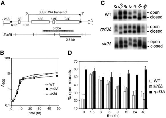
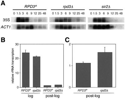
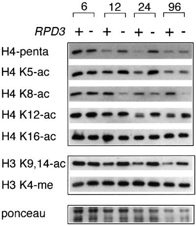
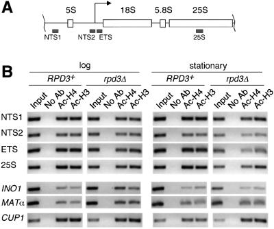
rRNA transcription in Saccharomyces cerevisiae is performed by RNA polymerase I and regulated by changes in growth conditions. During log phase, approximately 50% of the ribosomal DNA (rDNA) genes in each cell are transcribed and maintained in an open, psoralen-accessible conformation. During stationary phase, the percentage of open rDNA genes is greatly reduced. In this study we found that the Rpd3 histone deacetylase was required to inactivate (close) individual rDNA genes as cells entered stationary phase. Even though approximately 50% of the rDNA genes remained open during stationary phase in rpd3Delta mutants, overall rRNA synthesis was still reduced. Using electron microscopy of Miller chromatin spreads, we found that the number of RNA polymerases transcribing each open gene in the rpd3Delta mutant was significantly reduced when cells grew past log phase. Bulk levels of histone H3 and H4 acetylation were reduced during stationary phase in an RPD3-dependent manner. However, histone H3 and H4 acetylation was not significantly altered at the rDNA locus in an rpd3Delta mutant. Rpd3 therefore regulates the number of open rDNA repeats.


Figures

None
Fig. 1. Deletion of RPD3 prevents inactivation of rDNA repeats as cells exit log phase. (A) Schematic representation of the rDNA in S.cerevisiae, showing the initiation site and direction of 35S transcription by Pol I. The Pol III-transcribed 5S rRNA gene separates the non-transcribed spacer regions (NTS1 and NTS2). A Pol I enhancer element is located at the 3′ end of each 35S repeat (small black rectangles). The 2.8 kb EcoRI restriction fragment and probe are indicated. The boundaries of one rDNA repeat are indicated by vertical dashed lines. (B) Representative growth curve of yeast cultures used for the psoralen cross-linking assay. The strains used were JS311 (WT), JS490 (rpd3Δ) and JS218 (sir2Δ). (C) Gel retardation of actively transcribed rDNA genes through psoralen cross-linking. Aliquots harvested at the indicated time points were photoreacted with psoralen. Genomic DNA was digested with EcoRI and rDNA-specific fragments were detected using the 35S probe. For simplicity, only the 2.8 kb fragment is shown. The actively transcribed repeats (open) and inactive repeats (closed) are indicated on the right. (D) Quantitation of actively transcribed rDNA repeats. By PhosphorImager analysis, the percentage of the total rDNA repeats that were in the open chromatin conformation (slow migrating band) was calculated for each strain and time point. The average percentages from three independent experiments are shown in a bar graph along with standard deviation error bars.
None
Fig. 2. rRNA transcription from log phase and post-log phase yeast cultures. (A) Northern blot analysis of total RNA isolated from JS311 (RPD3+), JS490 (rpd3Δ) or JS566 (sir2Δ) cells that were harvested at the indicated time points after culture inoculation. Oligonucleotide probes were specific for the 35S rRNA precursor and the ACT1 mRNA. (BIn vivo transcriptional run-on assay for rRNA. Relative rRNA transcription amounts were plotted for log and post-log cells. (C) Enlarged graph of the post-log phase run-on data. Error bars are equal to the standard deviation from three independent experiments.
None
Fig. 3. Miller chromatin spread analysis of yeast rRNA transcription. (A) Typical clusters of rDNA genes from RPD3+ and rpd3Δ strains grown beyond log phase, approximating the diauxic shift. Examples of individual 35S genes are bracketed. Dark knobs at the end of transcripts represent rRNA processing machinery. (B) High magnification views of representative 35S genes from RPD3+ and rpd3Δ strains growing in log phase or post-log phase. (C) Graphical representation of the average number of polymerases that transcribe each active 35S gene in each strain and growth condition. Error bars represent the 99% confidence limits for the mean. The number (n) of genes analyzed for each condition is indicated.
None
Fig. 4. Bulk histone H3 and H4 acetylation status in stationary phase cells. JS311 (RPD3+) and JS490 (rpd3Δ) strains were grown from log into stationary phase. WHEs were analyzed by western blotting with a panel of histone H3- or H4-specific antibodies indicated on the left side of the panel. Hours of growth in YPD medium are indicated at the top. H3 K4-me antibody is specific for H3 methylated on lysine 4. This row represents a loading control, along with the Ponceau staining at the bottom.
None
Fig. 5. Histone H3 and H4 acetylation status of rDNA-associated chromatin in log and stationary phases. Specific DNA sequences that were associated with acetylated histone H3 or H4 were immunoprecipitated from RPD3+ (JS311) and rpd3Δ (JS490) WCEs. (A) The rDNA-specific sequences included NTS1, NTS2 (the Pol I promoter region), the 5′ ETS and the 25S transcribed region. The non-rDNA control sequences were the CUP1, MATα and INO1 genes. (B) PCR amplifications were performed with each primer pair. For the input lanes, 1/20 the amount of DNA was amplified for each rDNA location and CUP1 to remain in the linear range for the reaction. Ac-H4 represents the Penta H4 antibody. Ac-H3 represents the H3 K9/K14 acetyl antibody. The data shown are representative of four independent experiments. The fold differences between RPD3+ and rpd3Δ strains are shown in Table I.

References

    1. Aprikian P., Moorefield,B. and Reeder,R.H. (2001) New model for the yeast RNA polymerase I transcription cycle. Mol. Cell. Biol., 21, 4847–4855. - PMC - PubMed
    1. Ashrafi K., Sinclair,D., Gordon,J.I. and Guarente,L. (1999) Passage through stationary phase advances replicative aging in Saccharomyces cerevisiae. Proc. Natl Acad. Sci. USA, 96, 9100–9105. - PMC - PubMed
    1. Ausubel F.M., Brent,R., Kingston,R.E., Moore,D.D., Seidman,J.G., Smith,J.A. and Struhl,K. (2000) Current Protocols in Molecular Biology, Vol. 2. John Wiley & Sons, New York, NY.
    1. Bodem J., Dobreva,G., Hoffmann-Rohrer,U., Iben,S., Zentgraf,H., Delius,H., Vingron,M. and Grummt,I. (2000) TIF-IA, the factor mediating growth-dependent control of ribosomal RNA synthesis, is the mammalian homolog of yeast Rrn3p. EMBO Rep., 1, 171–175. - PMC - PubMed
    1. Braunstein M., Sobel,R.E., Allis,C.D., Turner,B.M. and Broach,J.R. (1996) Efficient transcriptional silencing in Saccharomyces cerevisiae requires a heterochromatin histone acetylation pattern. Mol. Cell. Biol., 16, 4349–4356. - PMC - PubMed

Publication types

MeSH terms