. 2002 Sep 16;21(18):4989-97.
doi: 10.1093/emboj/cdf458.

Visualization of parental HSV-1 genomes and replication compartments in association with ND10 in live infected cells

Affiliations

Visualization of parental HSV-1 genomes and replication compartments in association with ND10 in live infected cells

George Sourvinos et al. EMBO J. .

Abstract

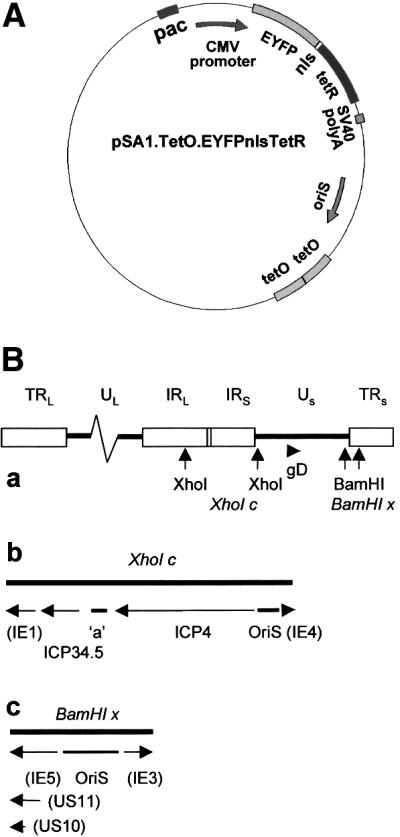
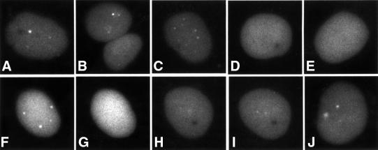
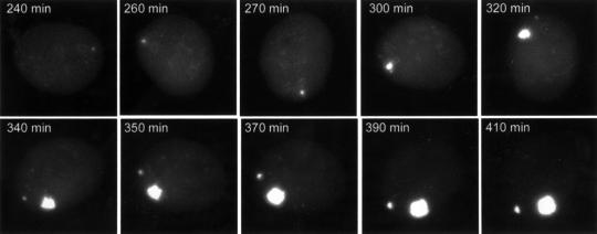
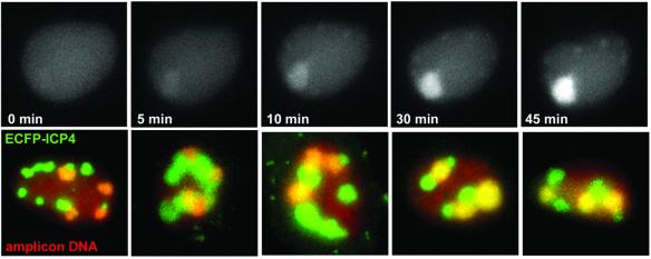
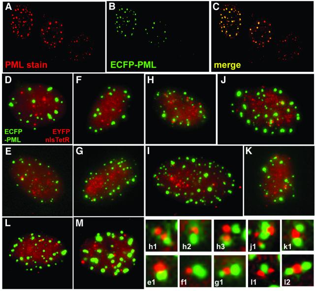
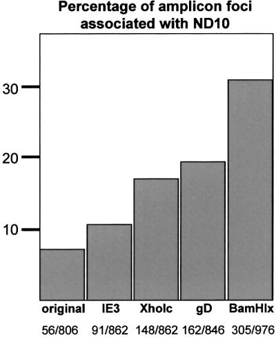
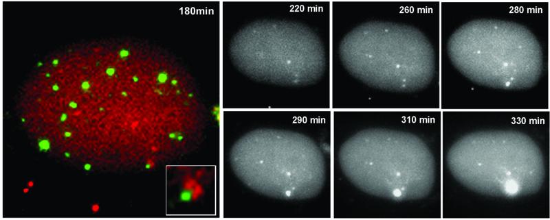
The relative location of active and repressed genes within the nucleus is becoming recognized as a significant factor in the control of gene expression. We have developed systems to visualize parental and replicated herpes simplex virus type 1 (HSV-1) amplicon genomes in association with PML nuclear bodies (ND10) in live cells. Plasmids containing viral replication and packaging signals, a gene expressing enhanced yellow fluorescent protein linked to the tetracycline repressor DNA binding domain and 14 copies of the tetracycline operator sequence were used to produce amplicon genomes packaged into normal viral particles. The frequency of the juxtaposition of viral genomes and ND10 was substantially increased by inclusion of an active HSV-1 Early gene transcription unit, indicating that the association is neither random nor passive. Furthermore, the ND10-associated genomes preferentially progressed to form viral replication compartments. Thus, active viral transcription contributes to the efficiency of viral genome association with ND10, and this in turn increases the probability that the genome will engage in active DNA replication. These studies in live cells provide a novel insight into virus-ND10 interactions and provide compelling visualization of their functional relevance.


Figures

None
Fig. 1. (A) Schematic representation of the basic amplicon plasmid pSA1.TetO.EYFPnlsTetR containing an HSV-1 OriS replication origin and packaging signals (pac). A CMV promoter drives expression of a fusion protein linking EYFP to a nls and the TetR DNA binding domain. Tandem reiterations of the 7-copy TetO sequence are also included. (B) Maps of the HSV-1 genome showing additional regions incorporated in the various amplicon plasmids: (a) the complete HSV-1 genome with the locations of the gD gene and the restriction sites bounding fragments XhoI c and BamHI x (the boxes depict the repeated sequences); (b) an expansion of the XhoI c region, with the locations of OriS, the included genes and the ‘a’ sequence marked; and (c) an expansion of the BamHI x region, with the locations of OriS and the included genes marked. In (b) and (c), the parentheses indicate that the promoter sequences, but not the complete gene, are included in the restriction fragment.
None
Fig. 2. Detection of parental amplicon genomes at early times after infection. Typical examples of EYFPnlsTetR amplicon-infected cells in the presence of DNA replication inhibitor ACG 90 min after infection. Parental amplicon genomes are detectable as distinct foci (AC) that are not observed in the presence of tetracycline (D and E). The dots formed in the presence of ACG (F) disappear within 10 min of adding tetracycline (G). Washing to remove the tetracycline allows recruitment of EYFPnlsTetR protein to the dots (H and I, same cell; J is another cell in the same sample after the tetracycline removal).
None
Fig. 3. The dynamics of developing amplicon replication compartments. Selected images of a time-lapse series of Vero cells co-infected with amplicon EYFPnlsTetR and wild-type HSV-1 helper virus are shown. The recruitment of the EYFPnlsTetR fusion protein into the amplicon replication compartment illustrates that replication is very rapid once it has been initiated. A second replication compartment starts developing at later stages of the infection. This particular cell exhibited rapid rotation during the early stages of the infection.
None
Fig. 4. (Upper panels) Amplicon replication compartments are not detected in the presence of tetracycline but become rapidly detectable after drug removal to allow binding of the EYFPnlsTetR protein onto replicated amplicon DNA. Cells were co-infected with EYFPnlsTetR amplicon and vECFP-ICP4 helper in the presence of tetracycline, and then the drug was removed 8 h after infection. An amplicon replication compartment became detectable only 5 min after removal of the drug and became rapidly brighter as pre-formed EYFPnlsTetR protein was sequestered onto pre-formed replicated amplicon DNA. (Lower panels) Five examples of cells in which both amplicon and helper virus replication compartments had developed between 5 and 6 h after infection. Both amplicon and helper virus replication centres are discrete, giving the concept of replication territories derived from individual parental genomes. The first two images were obtained using the gD amplicon, the third with the XhoI c derivative and the right-hand two with the BamHI x amplicon.
None
Fig. 5. (AC) Comparison of ND10 in uninfected Vero cells with those expressing ECFP–PML after a 16 h infection with baculovirus Ac.CMV.ECFP-PML. The left-hand two cells have been infected, whereas the right-hand cell is an uninfected control. (DM) Association of HSV-1 parental amplicon genomes with ND10 (all images were captured between 2 and 3 h after infection). The association between the EYFPnlsTetR amplicon genomes (red) and PML (green) in Vero cells was relatively rare (D and E). Similar frequencies of association were found after infection with the IE3 amplicon containing a model IE transcription unit (F and G). Parental amplicon genomes showed substantially increased juxtaposition with PML in cells infected with an amplicon containing an Early (gD) transcription cassette (H and I). Multiple associations between amplicon DNA and ND10 were also detected using the amplicon containing the 10.5 kb HSV-1 XhoI c fragment (J and K). A similar high incidence of association was observed when the BamHI x fragment containing the complete OriS replication origin region and IE, Early and Late promoters was included in the amplicon (L and M). Enlargement of examples of the juxtaposition of parental amplicon genomes and PML are shown in the lower right-hand panels (the lettering refers to the main panel from which the detail was selected). Apart from the typical association between single genome foci and single ND10 structures, examples of multiple interactions were also observed.
None
Fig. 6. Quantitation of the frequency of amplicon genome association with ND10. Over 800 individual amplicon foci in cells infected with each of the indicated amplicon stocks were scored for association with PML foci, shown as a percentage of the total. The actual numbers counted are shown in the bottom line.
None
Fig. 7. The development of an amplicon replication compartment in association with ND10. A cell containing several parental amplicon genomes (red) (co-infected with wild-type helper virus) was selected early during infection, and a double-labelled image was captured to show two genomes initially juxtaposed with a PML body (green) in the lower right-hand portion of the cell. The inset shows this region of the cell at a higher magnification. The greyscale panels show the EYFP-labelled amplicon genomes in the same cell during the course of the infection. A replication compartment developed from the genomes initially associated with the ND10 site, but not from other genomes that were not associated with PML foci. This is a typical example; several other cells were examined in other independent experiments, and in all cases replication compartments developed only from ND10-associated genomes.
None
Fig. 8. Preferential replication of amplicons with increased frequency of ND10 association. Stocks of amplicons expressing ECFPnlsTetR without additional sequences and EYFPnlsTetR containing the gD promoter cassette were prepared. Cells were infected with equalized amounts of the two amplicons either separately or together, and then the progeny supernatant virus was serially passaged twice more. DNA prepared from the cells of the three passages (as indicated) was cut with PstI and analysed by Southern blotting with markers derived from the amplicon plasmids. The 5.3 kb band identifies the ECFPnlsTetR amplicon, and the 3.5 and 0.54 kb bands identify the EYFPnlsTetR/gD amplicon. The 2.3 kb band is common to both. The EYFPnlsTetR/gD amplicon shows enhanced replication over the three passages.

References

    1. Bell P., Lieberman,P.M. and Maul,G.G. (2000) Lytic but not latent replication of Epstein–Barr virus is associated with PML and induces sequential release of nuclear domain 10 proteins. J. Virol., 74, 11800–11810. - PMC - PubMed
    1. Belmont A.S. (2001) Visualizing chromosome dynamics with GFP. Trends Cell Biol., 11, 250–257. - PubMed
    1. Belmont A.S. and Straight,A.F. (1998) In vivo visualization of chromosomes using lac operator–repressor binding. Trends Cell Biol., 8, 121–124. - PubMed
    1. Boyce F.M. and Bucher,N.L. (1996) Baculovirus-mediated gene transfer into mammalian cells. Proc. Natl Acad. Sci. USA, 93, 2348–2352. - PMC - PubMed
    1. Cockell M. and Gasser,S.M. (1999) Nuclear compartments and gene regulation. Curr. Opin. Genet. Dev., 9, 199–205. - PubMed

Publication types