

Flat and depressed colorectal tumours in a southern Swedish population: a prospective chromoendoscopic and histopathological study
- PMID: 12235079
- PMCID: PMC1773398
- DOI: 10.1136/gut.51.4.550
Flat and depressed colorectal tumours in a southern Swedish population: a prospective chromoendoscopic and histopathological study
Abstract
Background: Flat and depressed colorectal tumours are common in Japan but are very rare or non-existent in Western countries.
Aims: To study the occurrence of flat colorectal tumours in a southern Swedish population.
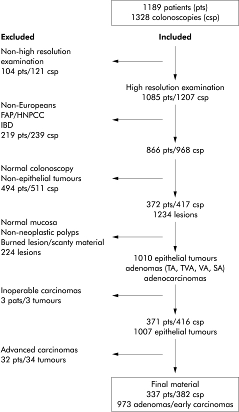
Methods: In this prospective study, 371 consecutive European patients were examined by high resolution video colonoscopy combined with chromoendoscopy. The nature of the lesions was determined by histopathological examination.
Results: A total of 973 tumours were found; 907 (93.2%) were protruding and 66 (6.8%) were flat or depressed. Of the flat/depressed tumours, five (7.7%) were early adenocarcinomas infiltrating the submucosa. Eleven carcinomas (1.2%) were found among protruding tumours. High grade dysplasia was observed in 18% (n=11) of flat/depressed adenomas in contrast with 7.3% (n=65) of protruding adenomas, and occurred in smaller flat/depressed tumours compared with protruding ones (mean diameter 8 mm v 23 mm, respectively). Furthermore, high grade dysplasia was significantly more common in flat elevated tumours with central depression or in depressed adenomas (35.7%; 5/14) than in flat elevated adenomas (12.8%; 6/47).
Conclusion: Flat and depressed tumours exist in a Western population. Future studies should address whether or not chromoendoscopy with video colonoscopy is necessary in the search for flat colorectal neoplasms.
Figures
References
-
- Morson BC. Precancerous and early malignant lesions of the large intestine. Br J Surg 1968;55:725–31. - PubMed
-
- Fenoglio CM, Lane N. The anatomic precursor of colorectal carcinoma. Cancer 1974;34:819–23. - PubMed
-
- Muto T, Bussey HJR, Morson BC. The evolution of cancer of the colon and rectum. Cancer 1975;36:2251–70. - PubMed
-
- Spratt JS, Ackerman LV. Small primary adenocarcinomas of the colon and rectum. JAMA 1962;179:337–46. - PubMed
-
- Kuramuto S, Oohara T. Minute cancers arising de novo in the hyman large intestine. Cancer 1988;61:829–34. - PubMed
Publication types
MeSH terms
LinkOut - more resources
Full Text Sources
Medical