. 2002 Sep;110(6):851-60.
doi: 10.1172/JCI15318.

Glucose-induced beta cell production of IL-1beta contributes to glucotoxicity in human pancreatic islets

Affiliations

Glucose-induced beta cell production of IL-1beta contributes to glucotoxicity in human pancreatic islets

Kathrin Maedler et al. J Clin Invest. 2002 Sep.

Erratum in

Abstract

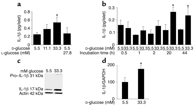
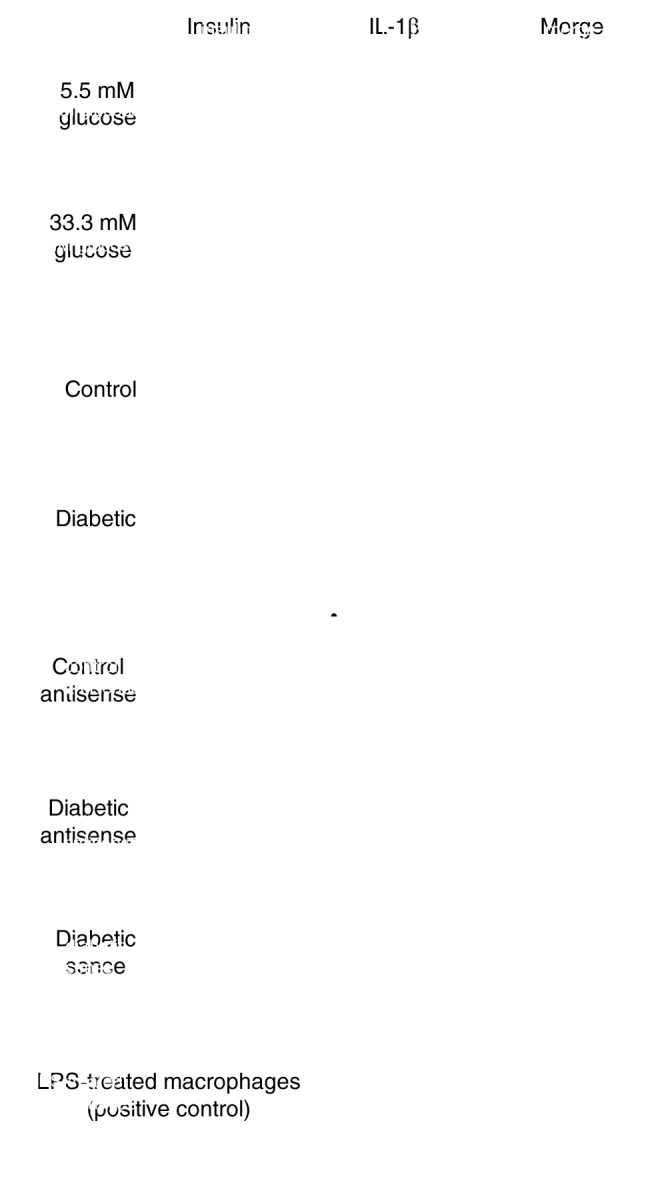
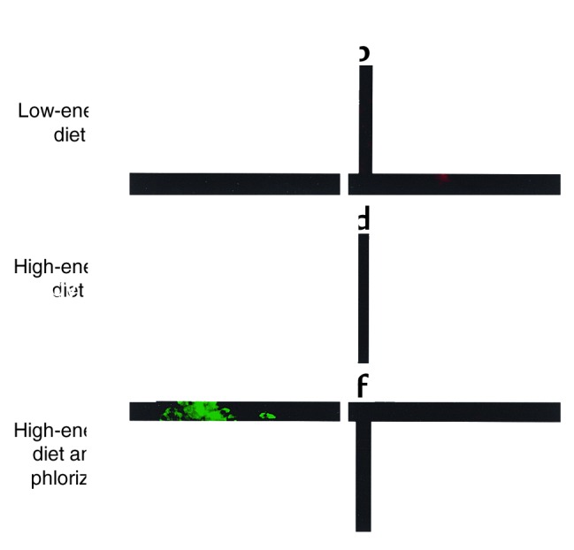
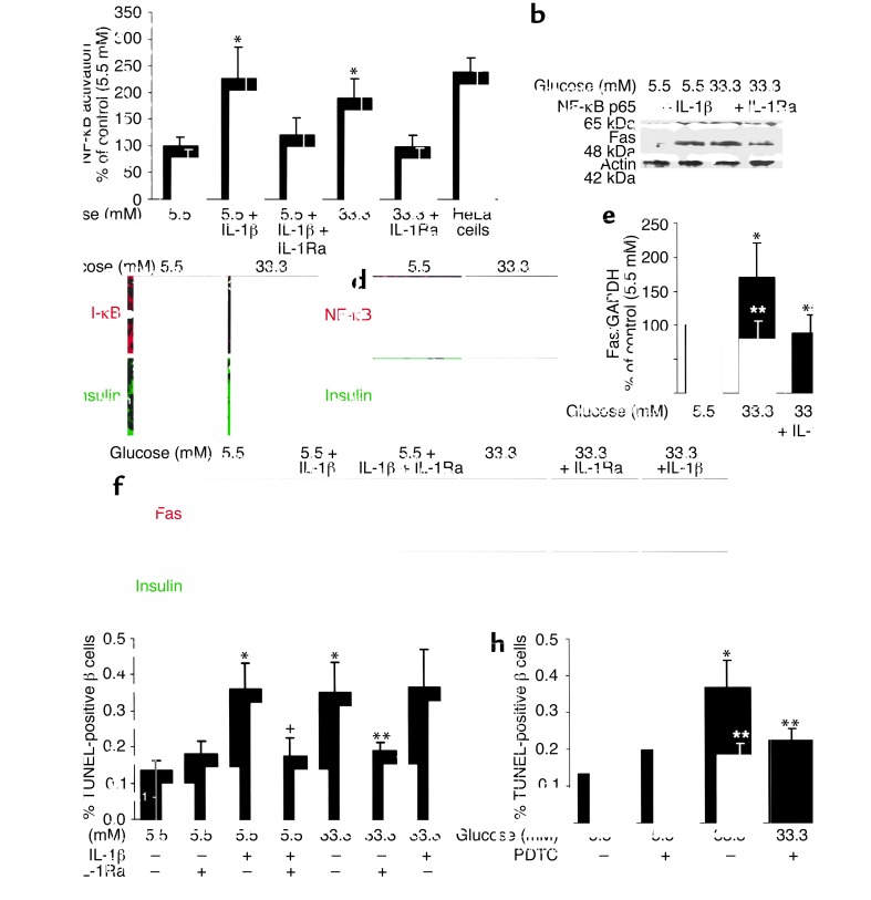
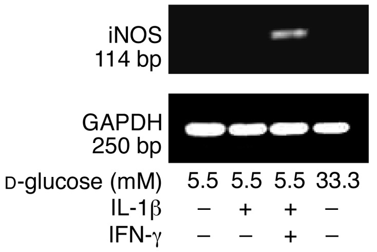
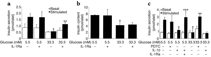
In type 2 diabetes, chronic hyperglycemia is suggested to be detrimental to pancreatic beta cells, causing impaired insulin secretion. IL-1beta is a proinflammatory cytokine acting during the autoimmune process of type 1 diabetes. IL-1beta inhibits beta cell function and promotes Fas-triggered apoptosis in part by activating the transcription factor NF-kappaB. Recently, we have shown that increased glucose concentrations also induce Fas expression and beta cell apoptosis in human islets. The aim of the present study was to test the hypothesis that IL-1beta may mediate the deleterious effects of high glucose on human beta cells. In vitro exposure of islets from nondiabetic organ donors to high glucose levels resulted in increased production and release of IL-1beta, followed by NF-kappaB activation, Fas upregulation, DNA fragmentation, and impaired beta cell function. The IL-1 receptor antagonist protected cultured human islets from these deleterious effects. beta cells themselves were identified as the islet cellular source of glucose-induced IL-1beta. In vivo, IL-1beta-producing beta cells were observed in pancreatic sections of type 2 diabetic patients but not in nondiabetic control subjects. Similarly, IL-1beta was induced in beta cells of the gerbil Psammomys obesus during development of diabetes. Treatment of the animals with phlorizin normalized plasma glucose and prevented beta cell expression of IL-1beta. These findings implicate an inflammatory process in the pathogenesis of glucotoxicity in type 2 diabetes and identify the IL-1beta/NF-kappaB pathway as a target to preserve beta cell mass and function in this condition.


Figures

Figure 1
Figure 1
Glucose induces IL-1β expression and release in human islets. (a) Secretion of IL-1β from human islets cultured on extracellular matrix–coated dishes for 4 days in 5.5, 11.1, or 33.3 mM D-glucose or in 5.5 mM D-glucose plus 27.8 mM L-glucose. Each bar represents the mean ± SEM of eight experiments from eight separate donors. *P < 0.01 compared with islets cultured in 5.5 mM glucose alone. (b) Secretion of IL-1β from human islets during 44 hours of culture in suspension with 5.5 or 33.3 mM D-glucose. Data were collected from four tubes per treatment in two separate experiments from two donors. Data are represented as mean ± SEM. *P < 0.01 compared with islets cultured in 5.5 mM glucose. (c) Immunoblotting of pro–IL-1β, IL-1β, and actin. Human islets cultured in suspension at 5.5 or 33.3 mM glucose were analyzed after 44 hours of incubation. One experiment of eleven (from eleven donors) is shown. In seven experiments, glucose induced IL-1β. In three experiments, IL-1β remained unchanged, and in one it was decreased. (d) RT-PCR detection and quantification of IL-1β mRNA expression. Total RNA was isolated from human islets cultured for 44 hours in medium containing 5.5 or 33.3 mM glucose. In the LightCycler quantitative PCR system, the level of IL-1β expression was normalized against GAPDH and the results were expressed as mRNA levels relative to control incubations at 5.5 mM. Results are presented as mean ± SEM for six independent experiments from six donors. *P < 0.05 relative to islets cultured in 5.5 mM glucose.
Figure 2
Figure 2
Expression of IL-1β by human β cells exposed to a diabetic milieu. Double immunostaining for IL-1β appears in red (b and d) and insulin in green (a and c) in human islets cultured on extracellular matrix–coated dishes and exposed for 4 days to media containing 5.5 mM glucose (a and b) or 33.3 mM glucose (c and d). Double immunostaining for IL-1β in red (f and h) and insulin in green (e and g) in tissue sections of pancreata from a nondiabetic patient (e and f) and from a patient with type 2 diabetes (g and h). In situ hybridization for IL-1β mRNA in red (j, l, and n) double immunostained for insulin in green (i, k, and m) in tissue sections of pancreata from a patient with type 2 diabetes (k, l, m, and n) with anti-sense probe (l) and with sense probe (negative control) (n), and from a nondiabetic patient (i and j) using anti-sense probe (j). Immunostaining for IL-1β in LPS-treated macrophages (positive control) (o). Magnification: ×250.
Figure 3
Figure 3
β cell expression of IL-1β during development of diabetes in Psammomys obesus. Double immunostaining for IL-1β in red (b, d, and f) and insulin in green (a, c, and e) in tissue sections of pancreata from a fasted Psammomys obesus on a low-energy diet (blood glucose 4 mM) (a and b), from an animal on a high-energy diet for 8 days without injection (blood glucose 13.6 mM) (c and d), and with injections of the glucopenic drug phlorizin (blood glucose 5.6 mM) (e and f). Magnification: ×250.
Figure 4
Figure 4
Glucose decreases β cell I-κB expression and induces IL-1β–meditated NF-κB activation, Fas expression, and DNA fragmentation. (a) Relative NF-κB activity. Human islets were cultured in suspension for 44 hours in 5.5 or 33.3 mM glucose alone or in the presence of IL-1β, IL-1Ra, or both. HeLa cells stimulated with 5 ng/ml IL-1α were used as positive control. Each bar represents the mean ± SEM of three experiments from three separate donors. *P < 0.05 relative to islets cultured in 5.5 mM glucose alone. (b) Immunoblotting of NF-κB (p65), Fas, and actin. Human islets cultured in suspension at 5.5 or 33.3 mM glucose with and without IL-1β or IL-1Ra were analyzed after 44 hours of incubation. The antibodies were blotted on the same membrane after stripping. One of three experiments from three donors is shown. Each experiment displayed similar results. (c) Double immunostaining for I-κB in red (boxes 1 and 3) and insulin in green (boxes 2 and 4) in sections of cultured human islets exposed for 44 hours to media containing 5.5 mM (boxes 1 and 2) or 33.3 mM glucose (boxes 3 and 4). (d) Double immunostaining for NF-κB (p65) in red (boxes 1 and 3) and insulin in green (boxes 2 and 4) in human islets exposed for 44 hours to media containing 5.5 mM (boxes 1 and 2) or 33.3 mM glucose (boxes 3 and 4). The arrows mark β cell nuclei stained positive for NF-κB. Magnification: ×750. (e) RT-PCR detection and quantification of Fas mRNA expression. Total RNA was isolated from human islets cultured for 44 hours in medium containing 5.5 or 33.3 mM glucose alone or in the presence of IL-1Ra. In the LightCycler quantitative PCR system, the level of Fas expression was normalized against GAPDH, and the results were expressed as mRNA levels relative to control incubations at 5.5 mM. Results are shown as mean ± SEM of six independent experiments from six donors. *P < 0.05 compared with islets cultured in 5.5 mM glucose alone. **P < 0.05 compared with islets cultured in 33.3 mM glucose. (f) Double immunostaining for Fas in red (boxes 1, 3, 5, 7, 9, and 11) and insulin in green (boxes 2, 4, 6, 8, 10, and 12) in human islets exposed for 4 days to media containing 5.5 mM glucose without IL-1β (boxes 1 and 2), with IL-1β alone (boxes 3 and 4), or with IL-1Ra (boxes 5 and 6) or 33.3 mM glucose without (boxes 7 and 8) and with IL-1Ra (boxes 9 and 10) or IL-1β (boxes 11 and 12). Magnification: ×250. (g) Human islets were cultured for 4 days in 5.5 and 33.3 mM glucose alone or in the presence of IL-1β and/or IL-1Ra, or (h) with and without PDTC. Results are mean ± SEM of the percentage of TUNEL-positive β cells. The mean number of islets scored from each donor was 49 (range 35–63) for each treatment condition. Islets were isolated from five organ donors. *P < 0.01 relative to islets cultured in 5.5 mM glucose. **P < 0.01 relative to islets cultured in 33.3 mM glucose. +P < 0.01 relative to islets cultured in 5.5 mM glucose plus IL-1β.
Figure 5
Figure 5
Failure of glucose and IL-1β to induce iNOS mRNA expression in human islets. RT-PCR analysis of iNOS expression by islets cultured for 44 hours in 5.5 and 33.3 mM glucose or with IL-1β alone or in combination with IFN-γ (positive control). GAPDH was used as control. One of three experiments from three donors is shown.
Figure 6
Figure 6
IL-1Ra and PDTC restore glucose-stimulated insulin secretion in human islets exposed to high glucose. Islets were cultured on extracellular matrix–coated dishes for 4 days in 5.5 and 33.3 mM glucose (control) or with IL-1Ra, PDTC, and IL-1β in combination or individually. (a and c) Basal and stimulated insulin secretion during successive 1-hour incubations at 3.3 (basal) and 16.7 (stimulated) mM glucose following the 4-day culture period. (b) Insulin content. Data are represented as mean ± SEM of three experiments from three separate donors. In each experiment, the data were collected from three plates per treatment. *P < 0.01 compared with islets cultured in 5.5 mM glucose alone. **P < 0.01 compared with islets cultured in 33.3 mM glucose alone. ***P < 0.01 compared with islets cultured in 5.5 mM glucose plus IL-1β.

References

    1. Unger RH, Grundy S. Hyperglycaemia as an inducer as well as a consequence of impaired islet cell function and insulin resistance: implications for the management of diabetes. Diabetologia. 1985;28:119–121. - PubMed
    1. Kaiser N, Corcos AP, Sarel I, Cerasi E. Monolayer culture of adult rat pancreatic islets on extracellular matrix: modulation of B-cell function by chronic exposure to high glucose. Endocrinology. 1991;129:2067–2076. - PubMed
    1. Leahy JL, Cooper HE, Deal DA, Weir GC. Chronic hyperglycemia is associated with impaired glucose influence on insulin secretion. A study in normal rats using chronic in vivo glucose infusions. J Clin Invest. 1986;77:908–915. - PMC - PubMed
    1. Robertson RP. Type II diabetes, glucose “non-sense,” and islet desensitization. Diabetes. 1989;38:1501–1505. - PubMed
    1. Rossetti L, Giaccari A, DeFronzo RA. Glucose toxicity. Diabetes Care. 1990;13:610–630. - PubMed

Publication types

MeSH terms