Comparative Study
. 2002 Sep 16;158(6):1079-87.
doi: 10.1083/jcb.200202049. Epub 2002 Sep 16.

Beta-catenin-induced melanoma growth requires the downstream target Microphthalmia-associated transcription factor

Affiliations
Comparative Study

Beta-catenin-induced melanoma growth requires the downstream target Microphthalmia-associated transcription factor

Hans R Widlund et al. J Cell Biol. .

Abstract

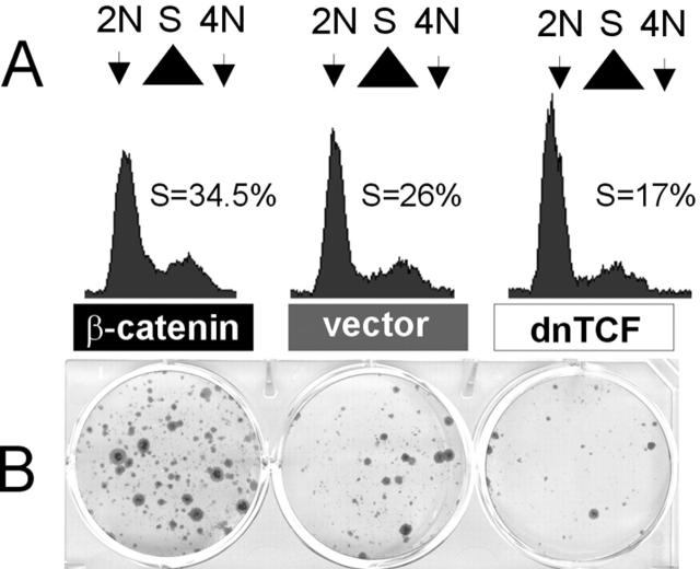
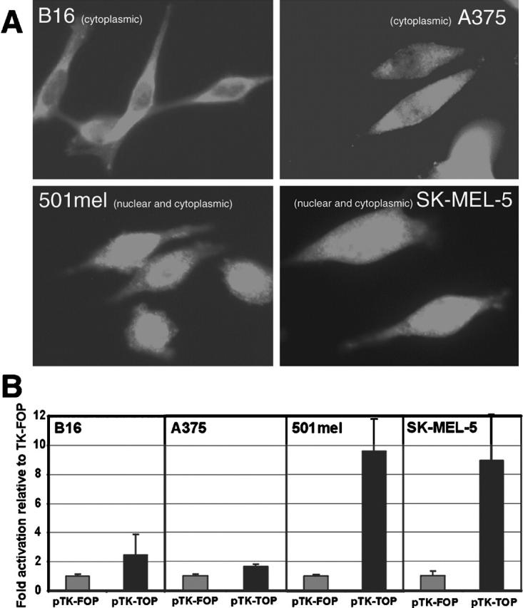
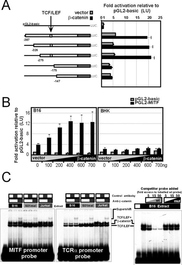
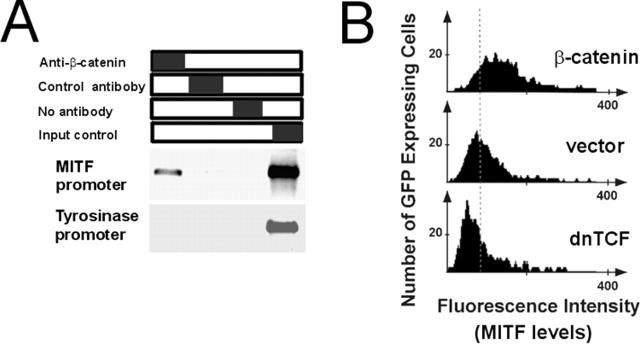
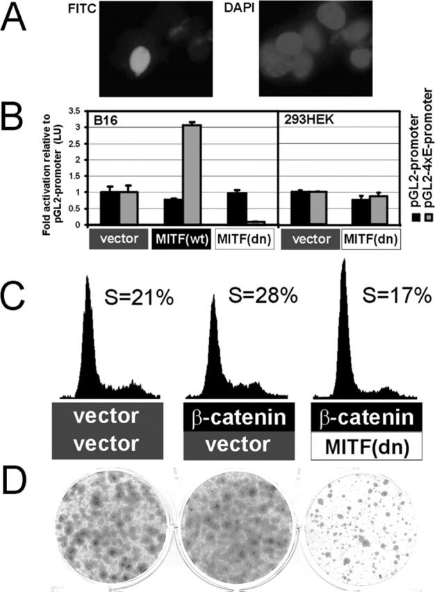
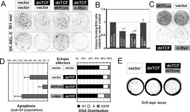
The transcription factor Microphthalmia-associated transcription factor (MITF) is a lineage-determination factor, which modulates melanocyte differentiation and pigmentation. MITF was recently shown to reside downstream of the canonical Wnt pathway during melanocyte differentiation from pluripotent neural crest cells in zebrafish as well as in mammalian melanocyte lineage cells. Although expression of many melanocytic/pigmentation markers is lost in human melanoma, MITF expression remains intact, even in unpigmented tumors, suggesting a role for MITF beyond its role in differentiation. A significant fraction of primary human melanomas exhibit deregulation (via aberrant nuclear accumulation) of beta-catenin, leading us to examine its role in melanoma growth and survival. Here, we show that beta-catenin is a potent mediator of growth for melanoma cells in a manner dependent on its downstream target MITF. Moreover, suppression of melanoma clonogenic growth by disruption of beta-catenin-T-cell transcription factor/LEF is rescued by constitutive MITF. This rescue occurs largely through a prosurvival mechanism. Thus, beta-catenin regulation of MITF expression represents a tissue-restricted pathway that significantly influences the growth and survival behavior of this notoriously treatment-resistant neoplasm.


Figures

Figure 1.
Figure 1.
Overexpression of β-catenin is proliferative and prosurvival in melanoma cells and dnTCF is inhibitory. (A) Cell cycle profiles of mouse B16 melanoma cells 48 h after transient transfection by indicated vectors and GFP as analyzed by propidium iodide staining and flow cytometry. (B) Representative colony-forming assays in mouse B16 melanoma cells.
Figure 2.
Figure 2.
Localization of endogenous β-catenin in melanoma cells and corresponding TCF/LEF transcriptional activity. (A) Melanoma cells (mouse B16 melanoma, human A375, human 501mel, and human SK-MEL-5) were assayed by immunofluorescence for the localization of β-catenin. As shown, B16 and A375 do not display aberrant nuclear accumulation of β-catenin in comparison with 501mel and SK-MEL-5, which display both cytoplasmic and nuclear β-catenin. (B) Bar graphs of the relative activity of TK-TOP, with optimal TCF/LEF binding sites and TK-FOP which carries mutated sites for TCF/LEF as an enhancer for the TK immediate early promoter driving luciferase.
Figure 3.
Figure 3.
β-catenin transactivates and binds the MITF promoter in vitro. (A) The TCF/LEF binding site is critical for β-catenin induced transactivation of the MITF promoter in B16 cells. (B) β-Catenin transactivation of the MITF promoter is tissue restricted, as in epithelial BHK cells the promoter is unresponsive to β-catenin. (C) Electrophoretic mobility shift assay showing that a complex formed from mouse B16, human 501mel, or Jurkat cell extracts on the MITF TCF/LEF probe can be specifically supershifted using an antibody against β-catenin. As a control, the same extracts were used to supershift a probe of the previously characterized TCRα TCF/LEF binding site. The complex visualized by EMSA is competed away by adding a wild-type MITF probe but not a mutant probe.
Figure 4.
Figure 4.
β-catenin is associated with the endogenous MITF promoter and modulates MITF expression levels. (A) In vivo association of β-catenin with the mouse MITF promoter by chromatin immunoprecipitation. The mouse tyrosinase promoter is a transcriptionally active negative control. (B) Ectopic expression of β-catenin leads to endogenous upregulation of MITF protein levels, and conversely, dnTCF downregulates MITF protein as quantitated by flow cytometry and analysis of transfected (gated GFP-positive) cells.
Figure 5.
Figure 5.
MITF(dn) blocks β-catenin–induced cell proliferation and growth in B16 melanoma cells. (A) Ectopic expression of an HA-tagged MITF(dn) in B16 melanoma cells is nuclear as visualized by FITC–anti-HA staining (compared with nuclear staining with DAPI in the same high power field). (B) MITF(dn) represses the activity of a 4 × E-box reporter that is transactivated by MITF(wt) in melanoma cells and this repression does not occur in the non-MITF expressing HEK293 cell line. (C and D) Expression of MITF(dn) abrogates β-catenin induced proliferation and growth in cell cycle analysis and clonogenic assay.
Figure 6.
Figure 6.
MITF can rescue growth suppression of dnTCF in melanoma cells. (A) Colony-forming assays in two human melanoma cell lines (501mel and SK-MEL-5) show rescue of clonogenic survival in the presence of dnTCF by wild-type MITF, whereas c-Myc is further growth inhibitory (B) Clonogenic survival in B16 melanoma demonstrates suppression by dnTCF and rescue by wild-type MITF, but not c-Myc (quantitative results are normalized to vector control). Experiment done in triplicate and quantitated results are graphed. Indicated with * is significant with P < 0.03 and ** significant with P < 0.01 as statistically calculated using Student's t test for unpaired samples. (C) Control colony formation assays in B16 cells showing growth suppressive effects of dnTCF and c-Myc in relation to MITF and vector control. (D) MITF rescues apoptosis induced by dnTCF but cannot relieve cell cycle inhibition in B16 melanoma cells. In contrast c-Myc bypasses dnTCF antiproliferative effects (S-phase repression), but significantly increases the apoptotic population. Apoptosis is measured as sub-G1 peak from propidium iodide staining. The quantitation was performed using ModFit v2.0 and represents values normalized to vector control as baseline. Averages from four independent experiments are presented (including error bars for standard deviation of mean). (E) Soft agar assay showing a representative example of macroscopic colonies of SK-MEL-5 cells 20 d after infection with the indicated retroviruses.

References

    1. Bentley, N.J., T. Eisen, and C.R. Goding. 1994. Melanocyte-specific expression of the human tyrosinase promoter: activation by the micropthalmia gene product and role of the initiator. Mol. Cell Biol. 14:7996–8006. - PMC - PubMed
    1. Bertolotto, C., P. Abbe, T.J. Hemesath, K. Bille, D.E. Fisher, J.P. Ortonne, and R. Ballotti. 1998. Microphthalmia gene product as a signal transducer in cAMP-induced differentiation of melanocytes. J. Cell Biol. 142:827–835. - PMC - PubMed
    1. Bienz, M., and H. Clevers. 2000. Linking colorectal cancer to Wnt signaling. Cell. 103:311–320. - PubMed
    1. Brault, V., R. Moore, S. Kutsch, M. Ishibashi, D.H. Rowitch, A.P. McMahon, L. Sommer, O. Boussadia, and R. Kemler. 2001. Inactivation of the β-catenin gene by Wnt-Cre-mediated deletion results in dramatic brain malformation and failure of craniofacial development. Development. 128:1253–1264. - PubMed
    1. Chang, K.L., and A.L. Folpe. 2001. Diagnostic utility of microphthalmia transcription factor in malignant melanoma and other tumors. Adv. Anat. Pathol. 8:273–275. - PubMed

Publication types

MeSH terms