Comparative Study
. 2002 Sep 16;158(6):1089-96.
doi: 10.1083/jcb.200203091. Epub 2002 Sep 16.

Involvement of TRPC in the abnormal calcium influx observed in dystrophic (mdx) mouse skeletal muscle fibers

Affiliations
Comparative Study

Involvement of TRPC in the abnormal calcium influx observed in dystrophic (mdx) mouse skeletal muscle fibers

Clarisse Vandebrouck et al. J Cell Biol. .

Abstract

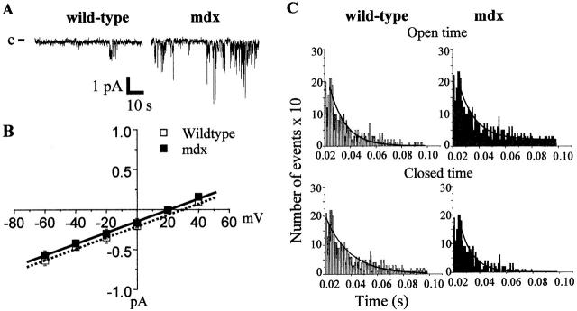
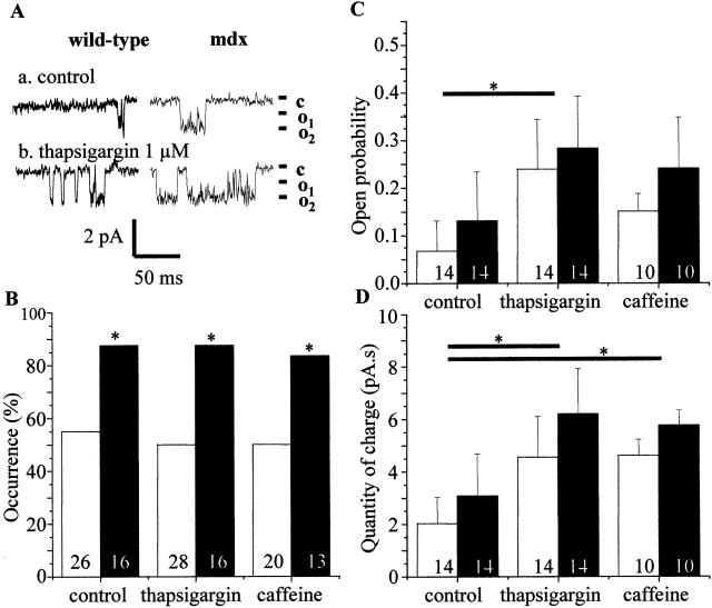
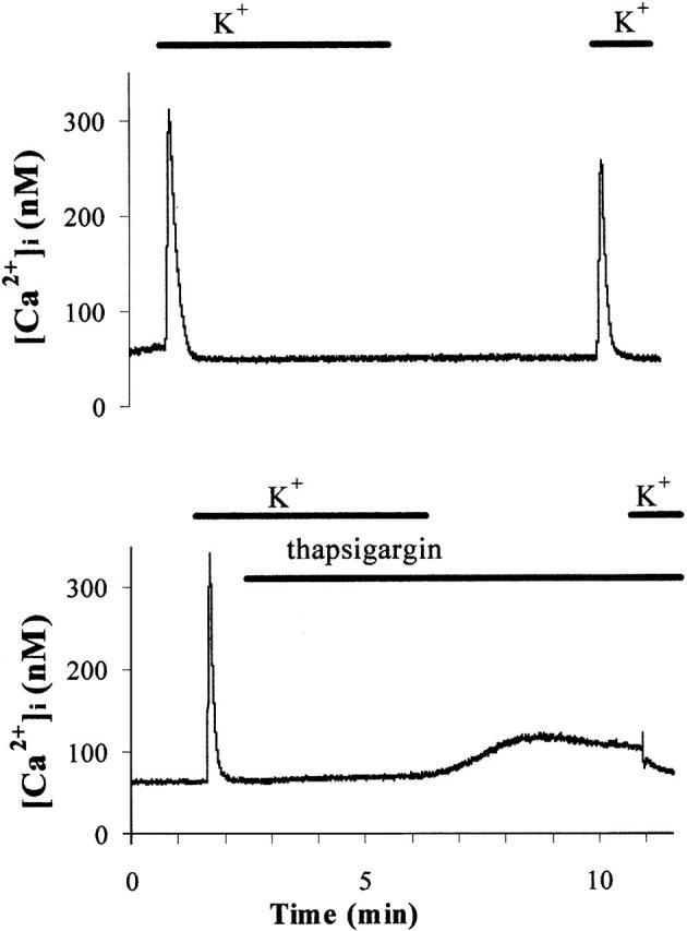
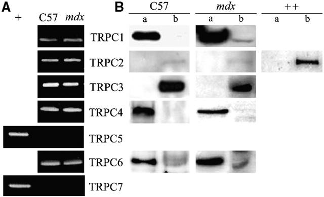
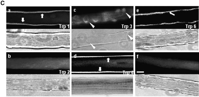
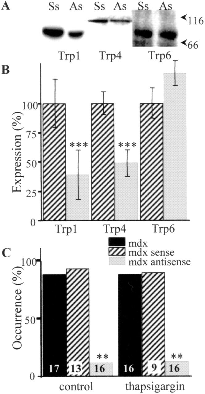
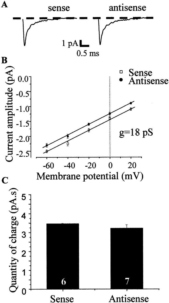
Duchenne muscular dystrophy results from the lack of dystrophin, a cytoskeletal protein associated with the inner surface membrane, in skeletal muscle. The absence of dystrophin induces an abnormal increase of sarcolemmal calcium influx through cationic channels in adult skeletal muscle fibers from dystrophic (mdx) mice. We observed that the activity of these channels was increased after depletion of the stores of calcium with thapsigargin or caffeine. By analogy with the situation observed in nonexcitable cells, we therefore hypothesized that these store-operated channels could belong to the transient receptor potential channel (TRPC) family. We measured the expression of TRPC isoforms in normal and mdx adult skeletal muscles fibers, and among the seven known isoforms, five were detected (TRPC1, 2, 3, 4, and 6) by RT-PCR. Western blot analysis and immunocytochemistry of normal and mdx muscle fibers demonstrated the localization of TRPC1, 4, and 6 proteins at the plasma membrane. Therefore, an antisense strategy was used to repress these TRPC isoforms. In parallel with the repression of the TRPCs, we observed that the occurrence of calcium leak channels was decreased to one tenth of its control value (patch-clamp technique), showing the involvement of TRPC in the abnormal calcium influx observed in dystrophic fibers.


Figures

Figure 1.
Figure 1.
Characterization of voltage-independent Ca channels in C57 and mdx fibers. (A) Examples of current traces (patch clamp; attached cell configuration). The closed state of the channel is marked by c. (B) Current- voltage relationships. (C) Distribution of open and closed times.
Figure 2.
Figure 2.
Thapsigargin and caffeine-activated single-channel currents in normal and mdx adult skeletal muscle fibers. (A) Original traces of single-channel inward currents recorded cell-attached membrane patch (at –60 mV) before (control) and after stimulation by 1 μM thapsigargin of wild-type and mdx fibers. The closed and open states of the channel are marked by c and o1 and o2, respectively. (B) Histogram showing the fraction of patches on normal (open bars) and mdx (filled bars) fibers with channel activity in control condition and after application of 1 μM thapsigargin or 10 mM caffeine. (C) Mean channel Po on normal C57 and mdx fibers. (D) Quantity of Ca charge in C57 and mdx fibers obtained by integrating recordings of cell-attached patches. The asterisk denotes significant difference (P < 0.05) between control and treated C57 fibers and between control and treated mdx fibers. The number of patches for each type of fibers in each condition is indicated in the histograms.
Figure 3.
Figure 3.
Representative examples of [Ca2 + ]i measurements in C57 muscle fibers. Fibers were incubated for 5 min in potassium aspartate (10 mM EGTA) solution in the presence or absence of 1 μM thapsigargin. Next, they were repolarized for 5 min in a Krebs solution (50 μM EGTA). Finally, depolarization in potassium aspartate solution induced a [Ca2+]i transient that was taken as an index of the amount of releasable Ca.
Figure 4.
Figure 4.
Expression and localization of TRPC isoforms. (A) Detection of the presence of TRPC mRNA by RT-PCR in C57 and mdx fibers. Brain (+) has been used as a positive control to prove the absence of TRPC5 and TRPC7. (B) Localization of endogenously expressed TRPC1, 2, 3, 4, and 6 in C57 and mdx fibers. Triton-soluble (a) or Triton-insoluble (b) proteins were separated on 8% SDS-PAGE, transferred onto PVDF membrane, and processed for Western blot analysis with specific pAbs. Sol 8 cells (++) have been used as a positive control for TRPC2. (C) Localization of Trp1, 2, 3, 4, and 6 proteins in mdx muscle fibers immunostained with specific antibodies (f, staining with the secondary antibody as a negative control). The corresponding light micrographs are presented for each case. Bar, 10 μm.
Figure 4.
Figure 4.
Expression and localization of TRPC isoforms. (A) Detection of the presence of TRPC mRNA by RT-PCR in C57 and mdx fibers. Brain (+) has been used as a positive control to prove the absence of TRPC5 and TRPC7. (B) Localization of endogenously expressed TRPC1, 2, 3, 4, and 6 in C57 and mdx fibers. Triton-soluble (a) or Triton-insoluble (b) proteins were separated on 8% SDS-PAGE, transferred onto PVDF membrane, and processed for Western blot analysis with specific pAbs. Sol 8 cells (++) have been used as a positive control for TRPC2. (C) Localization of Trp1, 2, 3, 4, and 6 proteins in mdx muscle fibers immunostained with specific antibodies (f, staining with the secondary antibody as a negative control). The corresponding light micrographs are presented for each case. Bar, 10 μm.
Figure 5.
Figure 5.
Repression of Trp in mdx fibers transfected with sense (Ss) or antisense (As) oligonucleotides. (A) Total transfected mdx fiber proteins were separated on 8% SDS-PAGE and blotted onto PVDF membrane. Western blots were performed with anti-Trp1, 4, and 6 pAbs. The standard molecular mass markers (kD) are indicated. (B) The intensities of the reactions were measured by integrated densitometry and were normalized for the protein load assayed by Coomassie blue staining. n = 6. ***, P < 0.0001. (C) Single-channel Ca activity in mdx fibers transfected with antisense or sense. Occurrence of channel activity in absence or presence of 1 μM thapsigargin. **, P < 0.001. The number of patches for each type of fibers in each condition is indicated in the histograms.
Figure 6.
Figure 6.
Sodium currents in mdx fibers transfected with sense or antisense. (A) Average currents constructed from 100 traces elicited in the same patches. (B) Single-channel current amplitudes plotted as a function of test potential for transfected mdx fibers. n ≥ 3. g, unitary conductance value. (C) Quantity of charge in mdx fibers (obtained by integrating recordings of cell-attached patches over a period of 120 s).

References

    1. Berridge, M.J. 1995. Capacitative calcium entry. Biochem. J. 312:1–11. - PMC - PubMed
    1. Bertorini, T.E., S.K. Bhattacharya, G.M. Palmieri, C.M. Chesney, D. Pifer, and B. Baker. 1982. Muscle calcium and magnesium content in Duchenne muscular dystrophy. Neurology. 32:1088–1092. - PubMed
    1. Boulay, G., D.M. Brown, N. Qin, M. Jiang, A. Dietrich, M.X. Zhu, Z. Chen, M. Birnbaumer, K. Mikoshiba, and L. Birnbaumer. 1999. Modulation of Ca2+ entry by polypeptides of the inositol 1,4, 5-trisphosphate receptor (IP3R) that bind transient receptor potential (TRP): evidence for roles of TRP and IP3R in store depletion-activated Ca2+ entry. Proc. Natl. Acad. Sci. USA. 96:14955–14960. - PMC - PubMed
    1. Clapham, D.E., L.W. Runnels, and C. Strubing. 2001. The TRP ion channel family. Nat. Rev. Neurosci. 2:387–396. - PubMed
    1. Collet, C., B. Allard, Y. Tourneur, and V. Jacquemond. 1999. Intracellular calcium signals measured with indo-1 in isolated skeletal muscle fibres from control and mdx mice. J. Physiol. 520:417–429. - PMC - PubMed

Publication types

MeSH terms