. 2002 Nov 15;277(46):44292-9.
doi: 10.1074/jbc.M205440200. Epub 2002 Sep 15.

Interaction of HCF-1 with a cellular nuclear export factor

Affiliations

Interaction of HCF-1 with a cellular nuclear export factor

Shahana S Mahajan et al. J Biol Chem. .

Abstract

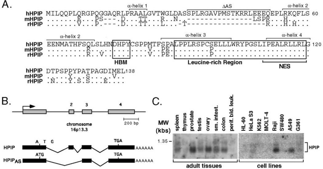
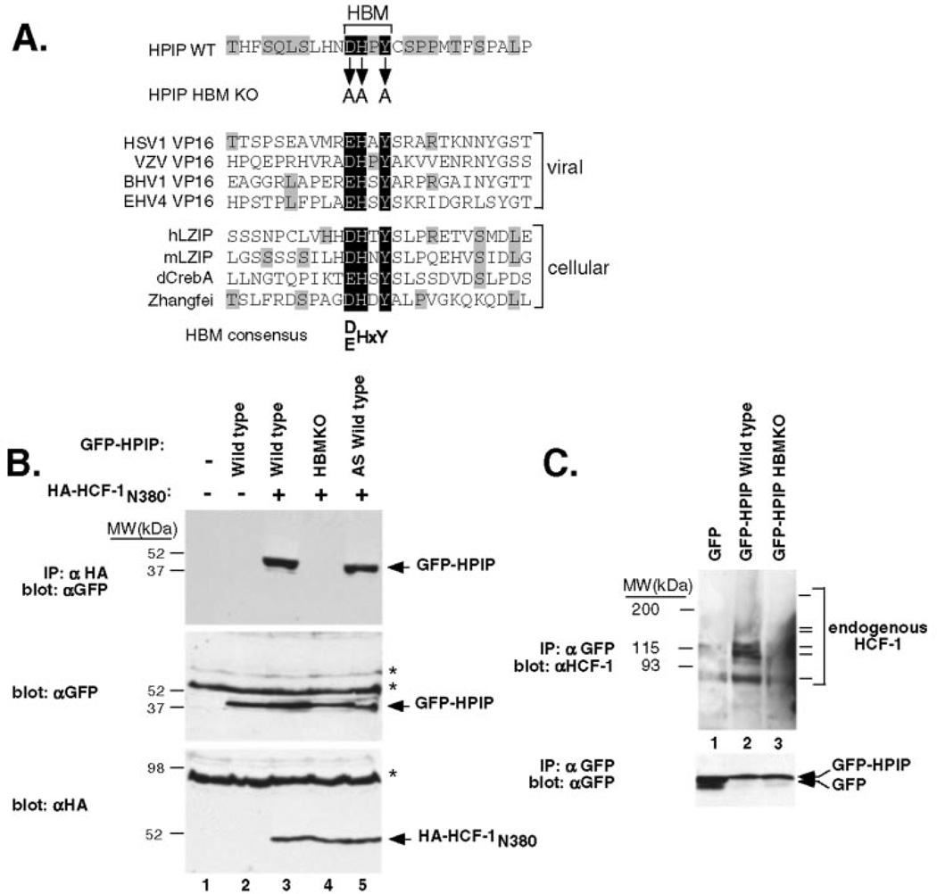
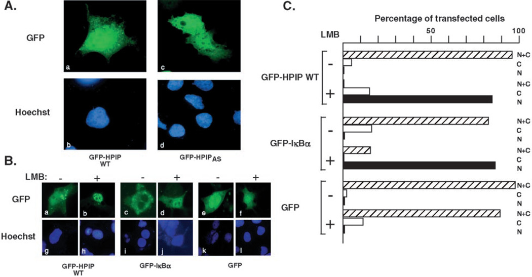
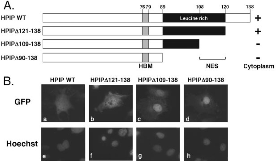
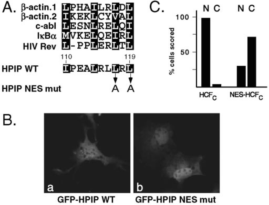
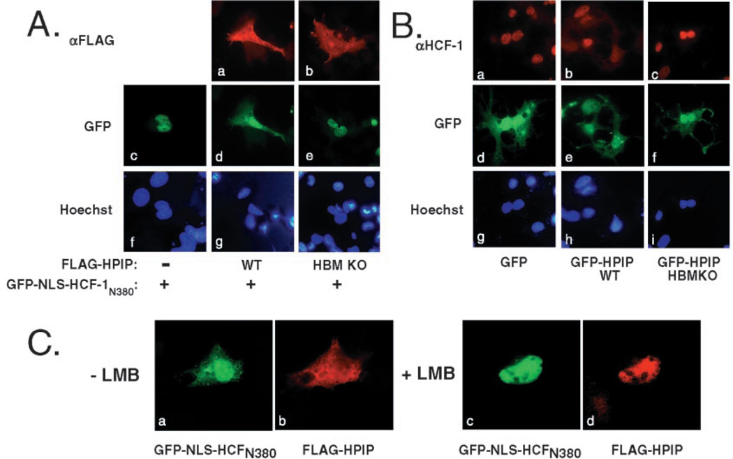
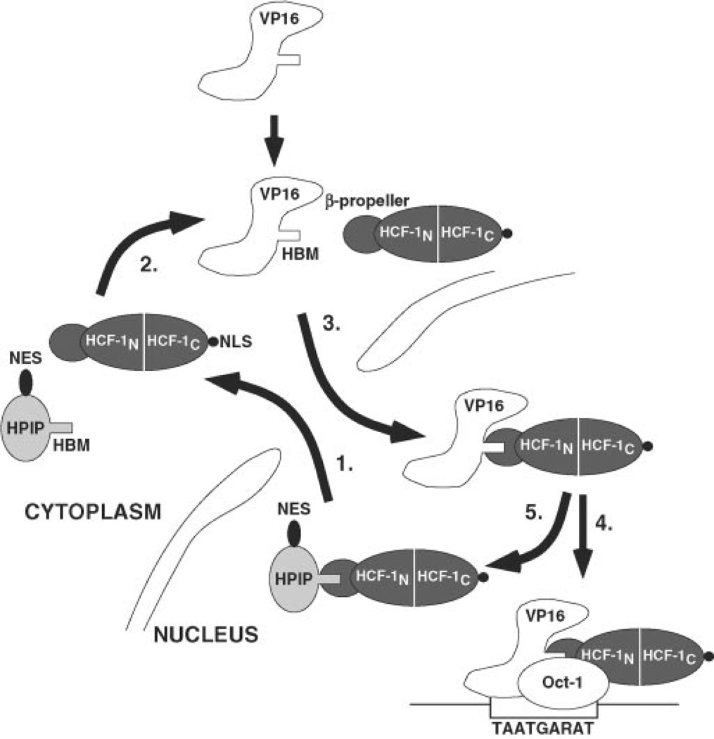
HCF-1 is a cellular protein required by VP16 to activate the herpes simplex virus (HSV) immediate-early genes. VP16 is a component of the viral tegument and, after release into the cell, binds to HCF-1 and translocates to the nucleus to form a complex with the POU domain protein Oct-1 and a VP16-responsive DNA sequence. This VP16-induced complex boosts transcription of the viral immediate-early genes and initiates lytic replication. In uninfected cells, HCF-1 functions as a coactivator for the cellular transcription factors LZIP and GABP and also plays an essential role in cell proliferation. VP16 and LZIP share a tetrapeptide HCF-binding motif recognized by the beta-propeller domain of HCF-1. Here we describe a new cellular HCF-1 beta-propeller domain binding protein, termed HPIP, which contains a functional HCF-binding motif and a leucine-rich nuclear export sequence. We show that HPIP shuttles between the nucleus and cytoplasm in a CRM1-dependent manner and that overexpression of HPIP leads to accumulation of HCF-1 in the cytoplasm. These data suggest that HPIP regulates HCF-1 activity by modulating its subcellular localization. Furthermore, HPIP-mediated export may provide the pool of cytoplasmic HCF-1 required for import of virion-derived VP16 into the nucleus.


Figures

Fig. 1
Fig. 1. Characterization of HPIP cDNA clones
A, predicted amino acid sequences of human (h), mouse (m), and rat (r) HPIP. Identical residues are indicated by a dot, whereas introduced gaps are indicated with dashes. The HCF-binding motif or HBM (DHPY) and leucine-rich region are boxed. In human cells, alternative mRNA splicing removes 19 residues (indicated as ΔAS) that are not conserved in the rodent counterparts. The nucleotide and predicted protein sequences reported here have been deposited in the GenBank™ data base with accession number AY116892. B, the deduced exon-intron structure of human HPIP, indicating the origins of the alternative splice variant (HPIPAS), which skips exon 2. C, Northern blot analysis showing the distribution of HPIP mRNA expression in a variety of human primary tissues and cells lines. MW, molecular mass.
Fig. 2
Fig. 2. HPIP and HCF-1 associate in mammalian cells
A, alignment of the HPIP sequence (black background) with the HBM regions of VP16-like proteins from herpes simplex virus-1 (HSV1), varicella-zoster virus (VZV), bovine herpesvirus-1 (BHV1), and equine herpesvirus-4 (EHV4) as well as the cellular HCF-1 interacting proteins human (hLZIP) and mouse LZIP (mLZIP), Drosophila dCREB-A/BBF2 (dCrebA), and human Zhangfei (15, 26, 54). Identical residues outside the core HBM are indicated with shading. B, 293T cells were cotransfected with expression plasmids encoding wild type or mutant versions of GFP-HPIP (3 µg) and HA-tagged HCF-1N380 (2 µg). The extracts were prepared and subject to coimmunoprecipitation using αHA antibody-coupled beads. Immunoprecipitates were resolved on a SDS-10% polyacrylamide gel and immunoblotted using an αGFP antibody (top panel). To monitor protein expression, extracts were blotted directly using the αGFP (middle panel) or αHA (bottom panel) antibodies. Nonspecific cross-reacting bands are indicated with asterisks. C, coimmunoprecipitation of endogenous HCF-1. The extracts were prepared from transfected 293T cells expressing GFP alone (lane 1), GFP-HPIP (lane 2), and GFP-HPIP HBM KO mutant (lane 3). After immunoprecipitation (IP) with αGFP antibody beads, the extracts were resolved by SDS-10% polyacrylamide gel electrophoresis and probed with αHCF-1 polyclonal sera (upper panel) or αGFP antibody (lower panel). The series of HCF-1 polypeptides detected by the αrHCF-H12 antibody (21) are indicated with bars. As reported previously, the higher molecular mass HCF-1300 and HCF-1150 polypeptides transfer poorly from 10% acrylamide gels and barely detected in this exposure.
Fig. 3
Fig. 3. HPIP shuttles between the nucleus and the cytoplasm
A, Cos-1 cells were transfected with expression plasmids (100 ng) encoding GFP-HPIP (panels a and c) and GFP-HPIPAS (panels b and d). The nuclei were counterstained with Hoechst dye 33258 (panels c and d). B, nuclear export of HPIP can be inhibited by LMB. Cos-1 cells expressing GFP-HPIP, GFP-IκBα, or GFP alone were seeded in duplicate, and 24 h post-transfection one set of samples was treated for 1 h with 20 ng/ml leptomycin B (+ LMB) or with vehicle alone (− LMB) and fixed for microscopy. The nuclei were counterstained with Hoechst 33258. C, quantitation of the experiment shown in B. At least 100 cells were scored for nuclear (N), cytoplasmic (C), and nuclear/cytoplasmic (N+C) distribution. The values are plotted as percentages of the total number of transfected cells examined.
Fig. 4
Fig. 4. The leucine-rich region is required for nuclear export
A, schematic showing HPIP truncations. Cytoplasmic (+) versus nuclear (−) accumulation is indicated. B, representative Cos-1 cells transfected with plasmids (100 ng) encoding GFP-HPIP WT (panels a and e), GFP-HPIP Δ121–138 (panels b and f), GFP-HPIP Δ109–138 (panels c and g), or GFP-HPIP Δ90–138 (panels d and h). After incubation for 24 h, the cells were fixed and analyzed. The nuclei were counterstained with Hoechst 33258 (panels e–h).
Fig. 5
Fig. 5. Identification of the HPIP nuclear export signal
A, alignment of the HPIP sequence (residues 110–119) with known NES from β-actin (55), c-abl (56), IκBα (57, 58), and HIV Rev (40, 59). The two-leucine residues in HPIP (Leu117 and Leu119) targeted for alanine substitution mutagenesis (HPIP NES mut) are indicated. B, Cos-1 cells were transfected with plasmids (100 ng) encoding GFP-HPIP or GFP-HPIP NES mut. C, quantitation of nuclear versus cytoplasmic staining (expressed as a percentage) of Cos-1 cells transfected with plasmids encoding GFP-HCF-1C (n = 112) or GFP-NES-HCF-1C (n = 107).
Fig. 6
Fig. 6. Expression of wild type HPIP promotes relocalization of HCF-1 to the cytoplasm
A, Cos-1 cells were transfected with plasmids (100 ng) expressing GFP-NLS-HCF-1N380 alone (panels c and f) or together with plasmids encoding FLAG-tagged HPIP WT (panels a, d, and g) or HPIP HBM KO (panels b, e, and h). After 24 h, the cells were fixed and probed using αFLAG primary antibody followed by a mouse IgG secondary antibody conjugated to Texas Red. The cells were stained with Hoechst dye to visualize the nuclei (panels f–h). B, as in A except that cells were transfected with plasmids encoding GFP alone (panels a, d, and g), GFP-HPIP WT (panels b, e, and h), and GFP-HPIP HBM KO (panels c, f, and i) and probed with an antibody against HCF-1 (panels a–c) to detect the endogenous HCF-1 protein. GFP (panels d–f) and DNA (panels g–i) were visualized by fluorescence. C, cytoplasmic accumulation of HCF-1 is inhibited by leptomycin B. Cos-1 cells were transfected with plasmids (100 ng) encoding GFP-NLS-HCF-1N380 and FLAG-tagged HPIP WT, reseeded in duplicate, and 24 h post-transfection treated for 1 h with vehicle alone (− LMB, panels a and b) or with 20 ng/ml leptomycin B (+ LMB, panels c and d).
Fig. 7
Fig. 7. Model for HPIP-mediated nucleocytoplasmic shuttling of HCF-1
Step 1, nuclear HCF-1 is exported to the cytoplasm through association with HPIP, which interacts with the N-terminal β-propeller domain (HCF-1N), which recognizes the HPIP HBM. The CRM-1 export protein (not illustrated) recognizes the NES in HPIP. Step 2, HCF-1 may dissociate from HPIP and return to the nucleus or return as complex with HPIP. Step 3, in herpes simplex virus-infected cells, VP16 is released into the cytosol together with other tegument components. The HCF-1 β-propeller domain interacts with the high affinity HBM of VP16, and the resulting complex is transported through the nuclear pore. Nuclear import of HCF-1 is mediated by its C-terminal NLS, which is recognized by the importin complex (11, 12). Once in the nucleoplasm, VP16, still complexed to HCF-1, can further associate with Oct-1 and the TAATGARAT sequence found in the viral immediate-early gene promoters. The resulting VP16-induced complex activates transcription by way of the potent C-terminal activation of VP16.

References

    1. Roizman B, Sears AE. In: Fields Virology. 3rd Ed. Fields BN, Knipe DM, Howley PM, editors. Vol. 2. Philadelphia, PA: Lippincott-Raven Publishers; 1996. pp. 2231–2295.
    1. O’Hare P. Semin. Virol. 1993;4:145–155.
    1. Wilson AC, Cleary MA, Lai J-S, LaMarco K, Peterson MG, Herr W. Cold Spring Harbor Symp. Quant. Biol. 1993;58:167–178. - PubMed
    1. Thompson CC, McKnight SL. Trends Genet. 1992;8:232–236.
    1. Stern S, Herr W. Genes Dev. 1991;5:2555–2566. - PubMed

Publication types

MeSH terms

Associated data