. 2002 Oct 1;99(20):13211-6.
doi: 10.1073/pnas.192314199. Epub 2002 Sep 16.

Experimental autoimmune encephalomyelitis mobilizes neural progenitors from the subventricular zone to undergo oligodendrogenesis in adult mice

Affiliations

Experimental autoimmune encephalomyelitis mobilizes neural progenitors from the subventricular zone to undergo oligodendrogenesis in adult mice

Nathalie Picard-Riera et al. Proc Natl Acad Sci U S A. .

Abstract

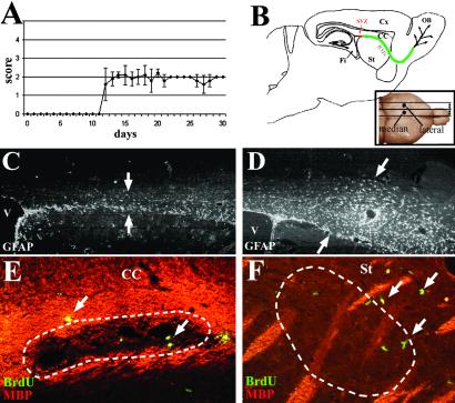
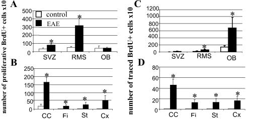
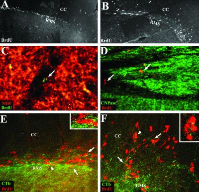
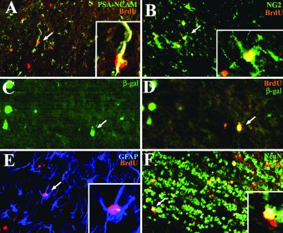
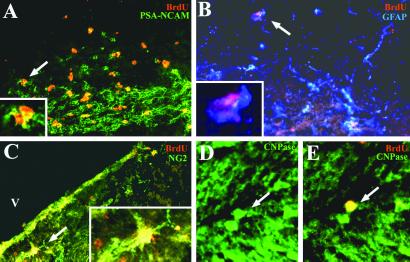
The destiny of the mitotically active cells of the subventricular zone (SVZ) in adult rodents is to migrate to the olfactory bulb, where they contribute to the replacement of granular and periglomerular neurons. However, these adult neural progenitors also can be mobilized in periventricular white matter and triggered to differentiate into astrocytes and oligodendrocytes in response to lysolecithin-induced demyelination. To mimic the environmental conditions of multiple sclerosis, we assessed the proliferation, migration, and differentiation potential of adult SVZ progenitor cells in response to experimental autoimmune encephalomyelitis (EAE) in mice. Inflammation and demyelination were observed in all mouse brains after EAE induction. EAE induced cell proliferation throughout the brain and especially within the lesions. Proliferating cells were neural progenitors, astrocytes, and oligodendrocyte precursors. EAE enhanced the migration of SVZ-derived neural progenitors to the olfactory bulb and triggered their mobilization in the periventricular white matter. The mobilized cells gave rise to neurons, astrocytes, and oligodendrocytes in the olfactory bulb but essentially to astrocytes and oligodendrocytes in the lesioned white matter. Our data indicate that the adult mouse SVZ is a source of newly generated oligodendrocytes and thus may contribute, along with oligodendrocyte precursors, to the replacement of oligodendrocytes in inflammatory demyelinating diseases of the central nervous system such as multiple sclerosis.


Figures

Figure 1
Figure 1
EAE mice displayed a moderate clinical course that began day 12 after induction. (A) Schematic representation of a sagittal brain section illustrating the localization of the SVZ and RMS to the OB and the various structures analyzed: striatum (St), fimbria (Fi), and cortex (Cx). (B Inset) Illustrates the localization of the median and the lateral areas studied. Immunohistochemistry 30 days p.i. shows that GFAP+ astrocytes were predominant in the RMS in the adjuvant control group (C, arrows) and expanded in the whole corpus callosum in the EAE mice (D, arrows). Double labeling for MBP (red) and BrdUrd (green) illustrates that proliferative cells (arrows) were detected in and ePose to demyelinated lesions (dotted lines) in the corpus callosum (E) and the striatum (F) of EAE mice. (C and D, ×44; E, ×178; F, ×88.)
Figure 2
Figure 2
Quantification of proliferative (A and B) and traced (C and D) cells, 30 days p.i., in control and EAE mice in structures where SVZ cells normally migrate such as the RMS and the OB (A and C) and in the white matter surrounding the SVZ such as the corpus callosum (CC), the striatum (St), the fimbria (Fi), and the cortex (Cx) (B and D). Data are expressed in number ± SD of BrdUrd+ cells per section ×10 and are averaged from the counts of three mice. Statistical analysis: Student's t test (*, P < 0.05).
Figure 3
Figure 3
Tracing of SVZ-derived cells. General view of BrdUrd-labeled cells migrating through the RMS 30 days p.i., in control (A) and EAE mice (B) (A and B are photomontages). In EAE mice, BrdUrd+ cells migrated extensively into the neighbor corpus callosum (CC). Some of the BrdUrd+ cells (arrows) reached demyelinating lesions in the corpus callosum (C) and in the fimbria (D) of induced mice. Double labeling for BrdUrd (red) and CTb (FITC), 7 days after intraventricular injection of CTb and i.p. injections of BrdUrd, demonstrated that BrdUrd-labeled cells originated from the SVZ, as seen in the RMS of control animals (arrows, E) and in the RMS and corpus callosum of EAE mice (arrows, F). Arrowheads identify cells magnified in Insets. [A and B, ×48; C, ×356; D and ×178; E and F, ×300, Insets (E and F): ×750.]
Figure 4
Figure 4
Characterization of traced cells in the OB of EAE mice 30 days p.i. Immunodetection of BrdUrd (red) and cell-specific markers (green or blue) shows that recruited cells were PSA-NCAM+ neural progenitors (A, arrow), NG2+ (B, arrow), and β-galactosidase+ (C and D, arrows) oligodendrocytes, GFAP+ astrocytes (E, arrow), and NeuN+ neurons (F, arrow). [A, B, E, and F, ×145; C and D, ×218; Insets (A, B, and E): ×410.]
Figure 5
Figure 5
Characterization of recruited cells in the corpus callosum of EAE mice 30 days p.i. Immunolabeling for BrdUrd (red) and cell-specific markers (green or blue) illustrates the presence of recruited cells that were PSA-NCAM+ neural progenitors (A, arrow), GFAP+ astrocytes (B, arrow), NG2+ oligodendrocytes precursors (C, arrow), and differentiated CNPase oligodendrocytes (D and E, arrows). [A–C, ×200; D and E, ×300; Insets (A–C): ×600.]

References

    1. Ffrench-Constant C, Raff M C. Nature (London) 1986;319:499–502. - PubMed
    1. Wolswijk G, Noble M. Development (Cambridge, UK) 1989;105:387–400. - PubMed
    1. Shi J, Marinovich A, Barres B A. J Neurosci. 1998;18:4627–4636. - PMC - PubMed
    1. Levine J M, Stincone F, Lee Y S. Glia. 1993;7:307–321. - PubMed
    1. Nishiyama A, Lin X H, Giese N, Heldin C H, Stallcup W B. J Neurosci Res. 1996;43:299–314. - PubMed

Publication types

Substances