. 2002 Oct 1;99(20):13009-13.
doi: 10.1073/pnas.202491499. Epub 2002 Sep 16.

T cells from the tumor microenvironment of patients with progressive myeloma can generate strong, tumor-specific cytolytic responses to autologous, tumor-loaded dendritic cells

Affiliations

T cells from the tumor microenvironment of patients with progressive myeloma can generate strong, tumor-specific cytolytic responses to autologous, tumor-loaded dendritic cells

Madhav V Dhodapkar et al. Proc Natl Acad Sci U S A. .

Abstract

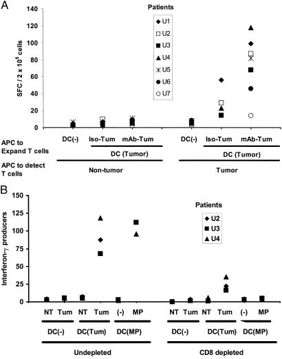
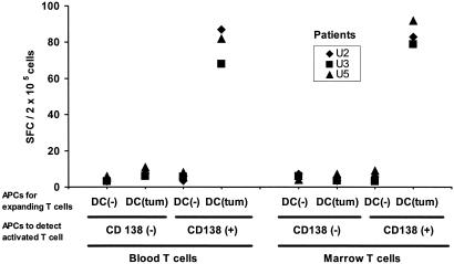
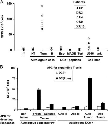
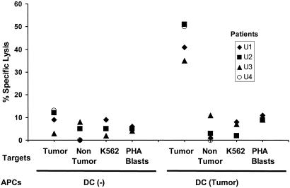
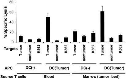
Most untreated cancer patients develop progressive tumors. We tested the capacity of T lymphocytes from patients with clinically progressive, multiple myeloma to develop killer function against fresh autologous tumor. In this malignancy, it is feasible to reproducibly evaluate freshly isolated tumor cells and T cells from the marrow tumor environment. When we did this with seven consecutive patients, with all clinical stages of disease, we did not detect reactivity to autologous cancer cells. However, both cytolytic and IFN-gamma-producing responses to autologous myeloma were generated in six of seven patients after stimulation ex vivo with dendritic cells that had processed autologous tumor cells. The antitumor effectors recognized fresh autologous tumor but not nontumor cells in the bone marrow, myeloma cell lines, dendritic cells loaded with tumor-derived Ig, or allogeneic tumor. Importantly, these CD8(+) effectors developed with similar efficiency by using T cells from both the blood and the bone marrow tumor environment. Therefore, even in the setting of clinical tumor progression, the tumor bed of myeloma patients contains T cells that can be activated readily by dendritic cells to kill primary autologous tumor.


Figures

Figure 1
Figure 1
Lack of detectable, myeloma-specific effector cells in freshly isolated T cells from the blood or marrow of myeloma patients. Freshly isolated T cells (105 cells per well) from the blood or bone marrow of an HLA A2-positive myeloma patient were cultured overnight (APC/responder ratio, 1:50) with fresh autologous tumor (CD138-positive) or nontumor (CD138-negative) fractions from the bone marrow (isolated by using magnetic beads) or autologous mature DCs pulsed for 2 h with several known HLA A2-restricted tumor antigen peptides (MAGE3-271–279 FLWGPRALV, NYESO-1-157–165 SLLMWITQC, and h-TERT1540 ILAKFLHWL, at 1 μg/ml), or control influenza peptide (MP58–66, GILGFLVFTL), or PHA (10 μg/ml) as a positive control. Antigen-specific IFN-γ-producing cells were quantified by using an ELISPOT assay. Data are representative of three HLA A2-positive patients tested.
Figure 2
Figure 2
Reliable expansion of tumor-specific effectors after stimulation with antigen-loaded DCs. (A) Antibody coating leads to enhanced generation of tumor-specific T cell responses. Monocyte-derived immature DCs from seven untreated patients with progressive myeloma were loaded with irradiated tumor cells, with or without prior opsonization with anti-syndecan-1 antibody (vs. isotype control) as described (7), or were left unpulsed. DCs then were matured by using cytokine mixture, and mature DCs were used to stimulate autologous blood T cells for 12–16 days, whereupon tumor-specific effectors were quantified against tumor (CD138-positive) or nontumor (CD138-negative) fractions from the marrow (APC/responder ratio, 1:50) by using an IFN-γ ELISPOT assay. (B) Effect of depletion of CD8+ T cells on reactivity of the tumor-specific, IFN-γ-producing cells. Blood T cells from an HLA A2-positive myeloma patient were stimulated for 12 days by using unpulsed DCs or DCs loaded with antibody-coated tumor cells or pulsed with an HLA A2-restricted influenza matrix peptide (MP), as described in Methods. After expansion, T cells were depleted of CD8+ fraction (or left undepleted) and tested for the presence of tumor-reactive or influenza peptide-specific T cells by using an ELISPOT assay, as described earlier (7). Data for influenza MP-specific effectors are per 5 × 10⁴ cells, and others are per 2 × 10⁵ cells. Tum, tumor; NT, nontumor fraction from autologous marrow.
Figure 3
Figure 3
Comparable expansion of tumor-specific effectors from blood and marrow T cells. Blood or marrow T cells were cultured with unpulsed or tumor cell-loaded DCs for 12–16 days. After expansion, the presence of tumor-reactive T cells was quantified with an ELISPOT assay by using autologous tumor (CD138+) or nontumor (CD138-ve) bone marrow cells as APCs, as described (7).
Figure 4
Figure 4
Nature of antigens by tumor-specific killer T cells. (A) T cells from HLA A2-positive patients expanded by using DCs loaded with antibody-coated tumor cells were tested for the recognition of autologous tumor or nontumor fractions from the marrow, purified autologous B cells, HLA A2.1-positive (U266) or negative (ark) myeloma cell lines, or HLA A2.1-restricted peptides derived from MAGE-3, h-TERT, or NY-ESO-1, using peptide-pulsed DCs as APCs (effector/APC ratio, 50:1). IFN-γ-producing cells (SFCs) were quantified in an ELISPOT assay. Data are shown as SFC/2 × 105 cells per well. (B) T cells (105 cells per well) from a patient with IgG myeloma expanded by using unpulsed DCs or DCs loaded with antibody-coated tumor cells were tested for the recognition of autologous CD138+ tumor [either fresh or cultured ex vivo in the presence of IL-6 (1,000 units/ml) and granulocyte/macrophage colony-stimulating factor (10 ng/ml) for 2 days] or nontumor cells (CD138-ve) from the bone marrow or autologous DCs pulsed with purified tumor-derived circulating Ig, allogeneic Ig, or DCs fed with autologous or allogeneic tumor cells (tumor/DC ratio, 1:1) as APCs. Effector cells were cultured overnight with APCs (effector/APC ratio, 50:1) and IFN-γ-producing cells (SFCs) quantified by an ELISPOT assay. Data are representative of experiments on two separate patients.
Figure 5
Figure 5
Generation of tumor-specific killer T cells in vitro. Blood T cells were cocultured with unpulsed or tumor cell-loaded DCs as in Fig. 2. T cells were tested for the ability to kill autologous tumor cells, nontumor fraction from the marrow, or K562 cells and autologous PHA blasts as controls at an effector-to-target ratio of 20:1.
Figure 6
Figure 6
Expansion of tumor-specific killers from blood or marrow T cells. T cells from blood and marrow T cells were expanded under conditions identical to those in Fig. 3 and tested for the ability to kill tumor or nontumor targets or K562 cells as controls. Data are representative of similar experiments on three separate patients.

References

    1. Tricot G. In: Hematology: Principles and Practice. Hoffman R, editor. New York: Churchill Livingstone; 2000. pp. 1398–1415.
    1. Cook G, Campbell J D. Blood Rev. 1999;13:151–162. - PubMed
    1. Heath W R, Carbone F R. Annu Rev Immunol. 2001;19:47–64. - PubMed
    1. Berard F, Blanco P, Davoust J, Neidhart-Berard E M, Nouri-Shirazi M, Taquet N, Rimoldi D, Cerottini J C, Banchereau J, Palucka A K. J Exp Med. 2000;192:1535–1544. - PMC - PubMed
    1. Wen Y J, Min R, Tricot G, Barlogie B, Yi Q. Blood. 2002;99:3280–3285. - PubMed

Publication types

MeSH terms