. 2002 Sep 15;30(18):3954-61.
doi: 10.1093/nar/gkf530.

G4 DNA unwinding by BLM and Sgs1p: substrate specificity and substrate-specific inhibition

Affiliations

G4 DNA unwinding by BLM and Sgs1p: substrate specificity and substrate-specific inhibition

Michael D Huber et al. Nucleic Acids Res. .

Abstract

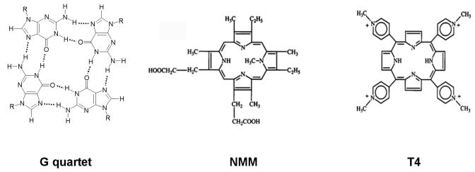
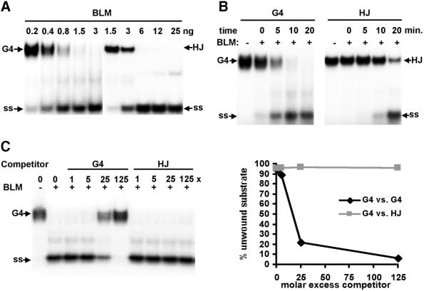
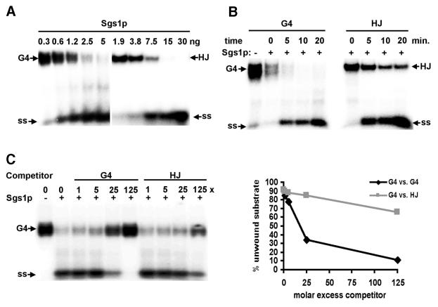
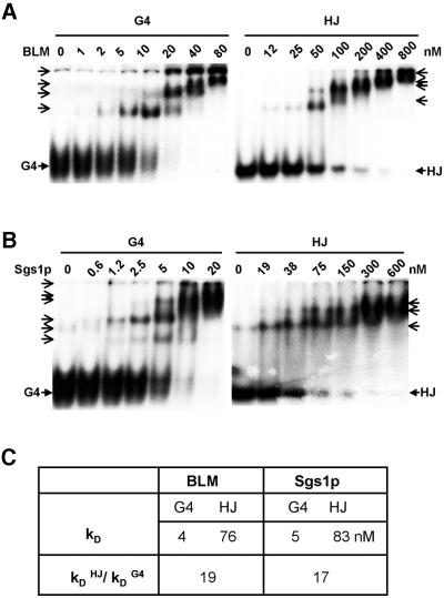
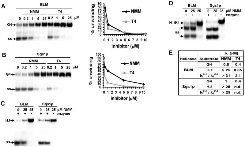
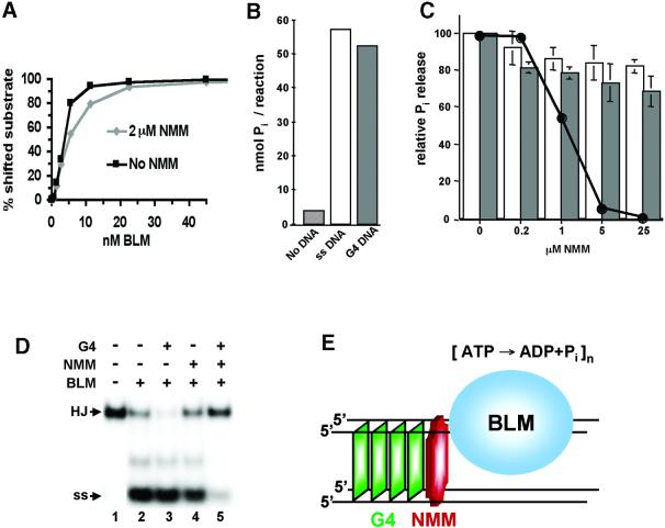
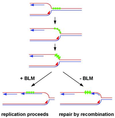
To understand the specific genetic instabilities associated with deficiencies in RecQ family helicases, we have studied the substrate preferences of two closely related members of this family, human BLM and Saccharomyces cerevisiae Sgs1p. Here we show that both BLM and Sgs1p preferentially unwind G4 DNA relative to Holliday junction substrates, and that substrate preference reflects binding affinity and maps to the conserved central helicase domain. We identify the porphyrin N-methyl mesoporphyrin IX (NMM) as a specific inhibitor of G4 DNA unwinding, and show that in the presence of NMM the helicase becomes trapped on the NMM-G4 DNA complex, consuming ATP but unable to unwind or dissociate. These results suggest that BLM and Sgs1p function proactively in replication to remove G4 DNA structures which would otherwise present obstacles to fork progression, rather than by promoting recombination to restart a fork that has stalled.


Figures

Figure 1
Figure 1
G4 DNA and porphyrins that bind it. A G quartet and porphyrin derivatives NMM and T4 (also referred to as TMPyP4 or H2TMPyP) are shown drawn to scale.
Figure 2
Figure 2
Preferential unwinding and binding of G4 DNA by BLM. (A) Native gel analysis of products of unwinding of 32P-labeled G4 DNA and HJ substrates by BLM. Amounts of enzyme per reaction are indicated above the autoradiograph. Arrows indicate G4 and HJ substrates and single-stranded unwinding products (ss). (B) Kinetics of unwinding of 32P-labeled G4 DNA and HJ, measured by native gel analysis. Products of reactions containing 5 ng of BLM were incubated at 37°C for the times indicated. (C) Unwinding of 32P-labeled G4 DNA in the presence of unlabeled G4 DNA or HJ competitor. Reactions containing 1 ng of BLM were incubated in the presence of the indicated molar excess of competitor and resolved on a native gel (left). Data are graphed on the right.
Figure 3
Figure 3
Preferential unwinding of G4 DNA by Sgs1p. (A) Native gel analysis of products of unwinding of 32P-labeled G4 DNA and HJ substrates by Sgs1p. Amounts of enzyme per reaction are indicated above the autoradiograph. Arrows indicate G4 and HJ substrates and single-stranded unwinding products (ss). (B) Kinetics of unwinding of 32P-labeled G4 DNA and HJ, measured by native gel analysis. Products of reactions containing 5 ng of Sgs1p were incubated at 37°C for the times indicated. (C) Unwinding of 32P-labeled G4 DNA in the presence of unlabeled G4 DNA or HJ. Reactions containing 5 ng of Sgs1p were incubated in the presence of the indicated molar excess of competitor and resolved on a native gel (left). Data are graphed on the right.
Figure 4
Figure 4
Preferential binding of BLM and Sgs1p to G4 DNA. (A) Gel mobility shift analysis of BLM binding to 32P-labeled G4 DNA and HJ at indicated protein concentrations. Positions of free G4 DNA and HJ are indicated by solid arrows. Open arrows indicate protein–DNA complexes. (B) Gel mobility shift analysis of Sgs1p binding to 32P-labeled G4 DNA and HJ at indicated protein concentrations. Symbols as in (A). (C) Summary of binding affinity, comparing kD for each helicase binding to G4 DNA and HJ, and showing relative affinities of binding to G4 DNA and HJ.
Figure 5
Figure 5
Inhibition of G4 DNA unwinding by NMM and T4. (A) Unwinding of 32P-labeled G4 DNA by BLM was assayed in the absence and presence of NMM and T4 at indicated concentrations, and products resolved on native gels (left). Data are graphed on the right. Symbols as in Figure 2. (B) Unwinding of 32P-labeled G4 DNA by Sgs1p, assayed and graphed as in (A). (C) Unwinding of 32P-labeled HJ by BLM and Sgs1p in the presence of NMM. (D) Unwinding of 32P-labeled partial duplex substrate H1/K1 by BLM and Sgs1p in the presence of NMM. (E) Summary of analysis of inhibition, comparing ki (concentration of inhibitor required to diminish unwinding activity 50%) for each enzyme and substrate.
Figure 6
Figure 6
Effect of NMM on G4 DNA binding and ATPase activity of BLM. (A) NMM does not alter BLM binding affinity for G4 DNA. Graph of results of gel mobility shift assays measuring binding of BLM to 32P-labeled G4 DNA, in the absence (squares) or presence (diamonds) of 2 µM NMM (2 × ki). (B) G4 DNA and ssDNA stimulate BLM ATPase activity. Pi release was measured in reactions containing no DNA (striped bar), 200 nM ssDNA (open bar) or 50 nM G4 DNA (shaded bar). (C) NMM does not alter BLM ATPase activity. Pi release was measured in reactions containing 200 nM ssDNA (open bars), or 50 nM G4 DNA (shaded bars). An unwinding inhibition curve (black line) derived from electrophoretic analysis of the same reactions is superimposed on the bar graphs. (D) The NMM–G4 DNA complex inhibits HJ unwinding. Unwinding of 32P-labeled HJ (1 pmol) by BLM (0.5 pmol) was assayed in the presence of unlabeled G4 DNA (0.2 pmol, lane 3); NMM (200 pmol, lane 4) or NMM–G4 DNA (lane 5). (E) Mechanism of inhibition of G4 unwinding by NMM. A helicase approaches the G4 structure from the 3′ end and encounters the masked face of the G4 stack. BLM continues to hydrolyze ATP but is unable to unwind or to dissociate from the substrate.
Figure 7
Figure 7
Model of the function of BLM or Sgs1p in replication. A replication fork approaches a G-rich region (green circles), and opening of the DNA duplex allows the G-rich strand to form G4 DNA (green triangles, third line). Either BLM or Sgs1p can recognize and unwind the G4 DNA, to produce a usable template strand (bottom line, left). In the absence of BLM or Sgs1p, G4 DNA persists, stalling replication. Replication restart by recombination results in the hyper-recombination characteristic of BLM- or SGS1-deficient cells.

References

    1. Ellis N.A., Groden,J., Ye,T.Z., Straughen,J., Lennon,D.J., Ciocci,S., Proytcheva,M. and German,J. (1995) The Bloom’s syndrome gene product is homologous to RecQ helicases. Cell, 83, 655–666. - PubMed
    1. Yu C.E., Oshima,J., Fu,Y.H., Wijsman,E.M., Hisama,F., Alisch,R., Matthews,S., Nakura,J., Miki,T., Ouais,S. et al. (1996) Positional cloning of the Werner’s syndrome gene. Science, 272, 258–262. - PubMed
    1. Kamath-Loeb A.S., Shen,J.C., Loeb,L.A. and Fry,M. (1998) Werner syndrome protein. II. Characterization of the integral 3′→5′ DNA exonuclease. J. Biol. Chem., 273, 34145–34150. - PubMed
    1. Kitao S., Shimamoto,A., Goto,M., Miller,R.W., Smithson,W.A., Lindor,N.M. and Furuichi,Y. (1999) Mutations in RECQL4 cause a subset of cases of Rothmund–Thomson syndrome. Nature Genet., 22, 82–84. - PubMed
    1. Sinclair D.A. and Guarente,L. (1997) Extrachromosomal rDNA circles—a cause of aging in yeast. Cell, 91, 1033–1042. - PubMed

Publication types