. 2002 Oct 1;99(20):12542-7.
doi: 10.1073/pnas.192457199. Epub 2002 Sep 17.

Gradients of substrate-bound laminin orient axonal specification of neurons

Affiliations

Gradients of substrate-bound laminin orient axonal specification of neurons

Stephan K W Dertinger et al. Proc Natl Acad Sci U S A. .

Abstract

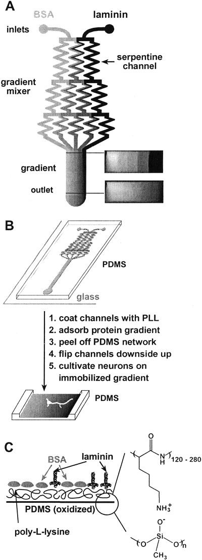
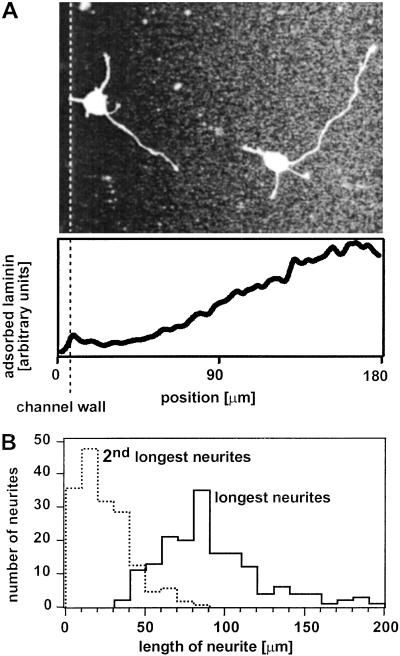
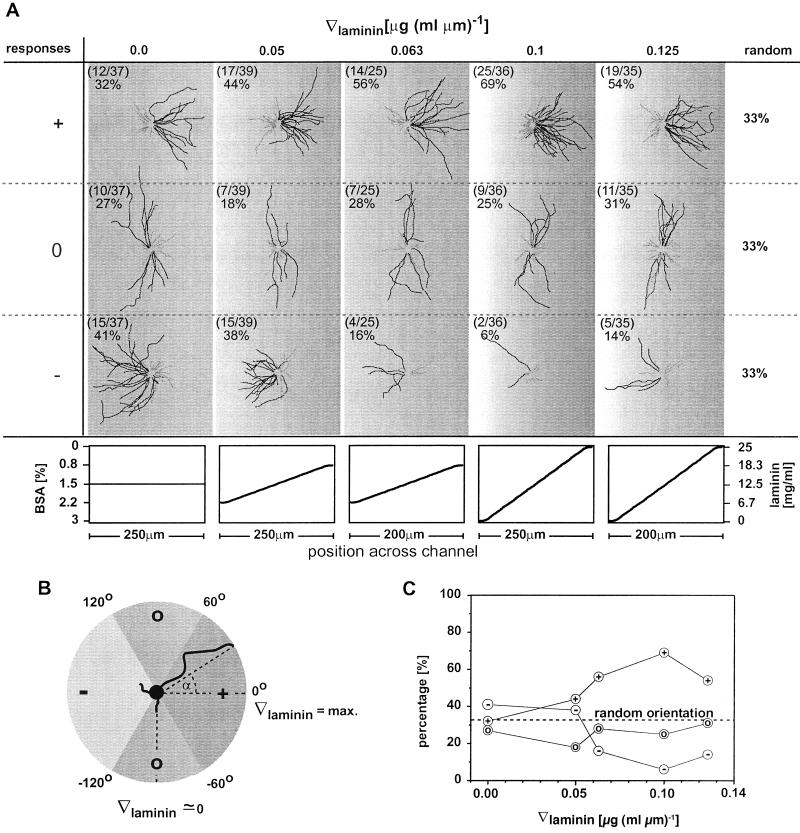
Little is known about the influence of substrate-bound gradients on neuronal development, since it has been difficult to fabricate gradients over the distances typically required for biological studies (a few hundred micrometers). This article demonstrates a generally applicable technique for the fabrication of substrate-bound gradients of proteins with complex shapes, using laminar flows in microchannels. Gradients that range from pure laminin to pure BSA were formed in solution by using a network of microchannels, and these proteins were allowed to adsorb onto a homogeneous layer of poly-l-lysine. Rat hippocampal neurons were cultivated on these substrate-bound gradients. Analysis of optical images of these neurons showed that axon specification is oriented in the direction of increasing surface density of laminin. Linear gradients in laminin adsorbed from a gradient in solution having a slope of nabla [laminin] > about 0.06 microg (ml.microm)(-1) (defined by dividing the change of concentration of laminin in solution over the distance of the gradient) orient axon specification, whereas those with nabla [laminin] < about 0.06 microg (ml.microm)(-1) have no effect.


Figures

Figure 1
Figure 1
(A) Schematic drawing of the design of a typical microfluidic network in PDMS that we used for the fabrication of immobilized gradients. Solutions of laminin and BSA were injected into the microfluidic network (inlets). Several streams, each carrying different concentrations of laminin and BSA, were generated in the gradient mixer and combined in a single channel to form a gradient perpendicular to the direction of the flow. The initial step profile created at the junction blurs slightly due to diffusion as the fluid travels downstream. The right-hand side of the diagram shows cross-sectional images of the concentration profile (using pure buffer and fluorescein, D = 5.0 × 10⁻⁶ cm2⋅s−1) of the flow visualized by using a confocal microscope. The shadows in the corners of the confocal images are due to the distortion of the light at the channel walls. (B) The diagram summarizes the steps from the fabrication of the substrate-bound gradient to the cultivation of neurons on the gradient immobilized on the substrate. A gradient was deposited on the floor of the channels by adsorption from the flowing stream. The network of microfluidic channels was removed, inverted, and placed in a culture dish. Neurons were plated on the floor of the channel in the region where the gradient had formed. (C) An idealized schematic diagram of the cross section of the surface composition of a substrate-bound gradient composed of laminin and BSA on PLL.
Figure 2
Figure 2
Rat hippocampal neurons preferentially extend their presumptive axon (their longest process) in the direction of increasing surface density of laminin. (A) A fluorescence micrograph of neurons that were fixed after 24 h in culture and immunostained for laminin (to visualize the substrate-bound gradient in laminin) and tubulin (to picture the microtubules of the neurons); staining for tubulin is commonly used to visualize the morphology of a neuron. Fluorescent images of tubulin and laminin have been superimposed. The shape of the immobilized gradient in laminin is shown in the graph below the micrograph. The fluorescent signal of the immunostained laminin has been used to measure the shape of the gradient. The slope of the laminin gradient in solution used to fabricate the substrate-bound gradient was ∇[laminin] = 0.125 μg (ml⋅μm)−1. The dotted line indicates the left wall of the channel. The walls of the channel and local bright spots distort the uniformity of the fluorescent intensities (e.g., on the left and right edges of the micrograph). The right wall of the channel is at 250 μm (not shown). (B) A histogram of the length of the longest and second longest process of all neurons that were included in the statistical analysis of axon orientation. After 24 h in culture, the longest process of a neuron is on average four times longer than the second longest process.
Figure 3
Figure 3
(A) Superimposed drawings of all processes of neurons grown on gradients of laminin on PLL. Neurons were grouped and superimposed according to the orientation of the tip of the axon (the longest process) after 24 h in culture. This analysis was done for each gradient slope separately (columns). The single axon of a neuron is drawn in black; the remaining short processes (dendrites) are drawn in gray. Neurons with axons extending in the direction of increasing laminin concentration are shown in the first row. Neurons with axons extending parallel to the gradient or in the direction of decreasing laminin concentration are shown in the second and third row, respectively. The shapes of the gradients in laminin and BSA in solution that were used to fabricate the substrate-bound gradient are given in the graphs below. The gradients in laminin and BSA were always formed on a homogeneous layer of PLL. In each drawing, the number of neurons in each classification (+, 0, or −) is listed with the total number of neurons grown on a particular gradient on the upper left corner. (B) Definition of the angle α of axonal process grown on a gradient in laminin. The sectors that formed the basis for the classification in (+)-, (0)-, and (−)-responding neurons are indicated. (C) Summary of the percentage of (+)-, (0)-, and (−)-responding neurons cultivated on gradients with different slopes in laminin.

References

    1. Tessier-Lavigne M, Goodman C S. Science. 1996;274:1123–1133. - PubMed
    1. Song H J, Poo M M. Curr Opin Neurobiol. 1999;9:355–363. - PubMed
    1. Baier H, Bonhoeffer F. Science. 1992;255:472–475. - PubMed
    1. Dertinger S K W, Chiu D, Jeon N L, Whitesides G M. Anal Chem. 2001;73:1240–1246.
    1. Dotti C G, Sullivan C A, Banker G A. J Neurosci. 1988;8:1454–1468. - PMC - PubMed

Publication types

LinkOut - more resources