. 2002 Oct 1;99(20):13073-8.
doi: 10.1073/pnas.192586699. Epub 2002 Sep 17.

Growth suppression of human carcinoma cells by reintroduction of the p300 coactivator

Affiliations

Growth suppression of human carcinoma cells by reintroduction of the p300 coactivator

Tamaki Suganuma et al. Proc Natl Acad Sci U S A. .

Abstract

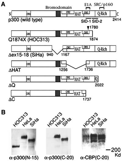
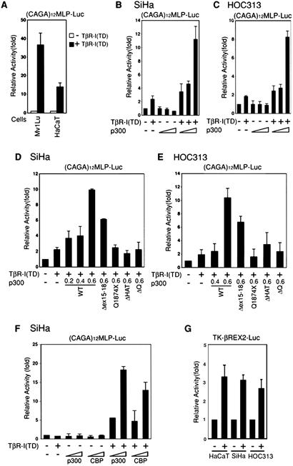
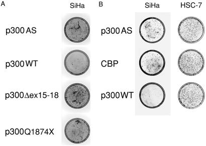
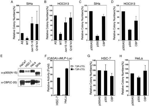
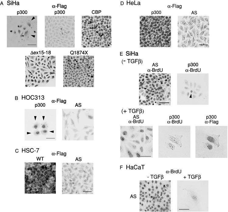
The p300 and closely related cAMP response element binding protein (CREB)-binding protein (CBP) acetyltransferases function as global transcriptional coactivators and play important roles in a broad spectrum of biological processes, including cell proliferation and differentiation. A role of p300/CBP in tumor suppression has been proposed from the fact that these coactivators are targeted by viral oncoproteins and that biallelic mutations of p300 have been identified in carcinomas. Here, we show that transcriptional response to the transforming growth factor beta (TGF-beta), an inhibitor of epithelial cell growth, was severely impaired in human carcinoma cell lines carrying p300 mutations accompanied by inactivation of the second allele, and that wild-type expression restored TGF-beta-dependent transcriptional activity. Furthermore, reintroduction of wild-type p300 suppressed the growth of p300-deficient carcinoma cells, whereas p300 did not inhibit the growth of carcinoma cells examined, which have no detectable alterations in p300 protein and retain the TGF-beta-dependent transcriptional response. In addition, tumor-derived mutants missing the bromodomain or glutamine-rich region, which are respectively important for chromatin interaction and coactivator activities, lost the suppressive activity. In contrast, CBP exhibited no or reduced ability to suppress the growth of p300-deficient carcinoma cells. These results provide experimental evidence to show that p300 acts as a suppressor of tumor cell growth and suggest a distinct role of p300 in suppression of epithelial tumors.


Figures

Figure 1
Figure 1
Mutations of p300 in HOC313 and SiHa cells. (A) Schematic diagram of wild type and p300 mutants. Functional domains of p300 protein are indicated: Cys/His-rich regions (CH1, CH2, and CH3), bromodomain, HAT domain (HAT), E1A-binding region (E1A), SRC/p160-binding region, Q-Rich, and Smad interaction domains (SID-1 and SID-2). Numbers indicate amino acids at boundaries. Position of mutation at codon 1780 is indicated by an arrowhead. (B) Alterations of p300 protein in HOC313 and SiHa cells. Western blotting of HeLa, HOC313, and SiHa cells. The blot was probed with α-p300 (N-15; Left), then reprobed for α-p300 (C-20; Center), and α-CBP (C-20; Right).
Figure 2
Figure 2
TGF-β-dependent transcriptional activity is restored by p300 in SiHa and HOC313 cells. (A) Mv1Lu and HaCaT cells were transfected with 0.3 μg of (CAGA)12MLP-Luc reporter, together with 0.1 μg of pTβR-I (TD). (B and C) SiHa (B) and HOC313 (C) cells were transfected with (CAGA)12MLP-Luc, together with pTβR-I (TD) as in A, and increasing amounts (0.2, 0.4, and 0.8 μg in B or 0.4, 0.6, and 0.8 μg in C) of p300. (D and E) Experiments were performed as described in B and C except that the indicated amounts (in μg) of wild-type or mutant p300 plasmids were used. (F) Experiments were performed as described in B except that 0.4 and 0.6 μg of p300 or CBP plasmids were used. (G) Response of SiHa and HOC313 cells to RAR activity. SiHa, HOC313, and HaCaT cells were transfected with 0.25 μg of pTK-βREX2-Luc reporter, together with a plasmid expressing RARα and then stimulated with 9cis-RA (10−6 M) 24 h before harvest. Results are shown as relative luciferase levels. Data represent the average of three independent experiments, each performed in duplicate.
Figure 3
Figure 3
Colony formation assay with SiHa and HSC-7 cells. Cells were stably transfected with 2.5 μg of control pDEFp300AS or indicated Flag-tagged p300 or CBP expression plasmids in a 35-mm plate, split into two 100-mm plates, and then cultured in media containing G418 for 3 wk. Cells were stained with Giemsa solution. (A and B) Photographs of plates showing colony formation assay of SiHa and HSC-7 cells.
Figure 4
Figure 4
Suppression of carcinoma cell growth by p300. Colony formation assays proceeded as in Fig. 3, and then the number of colonies was counted. Data represent the average of the number of colonies obtained from at least three independent experiments. (A and B) Suppression of colony formation by wild-type, but not mutant, p300 in SiHa (A) and HOC313 (B) cells. The mark at 100% represents 780 and 114 colonies per plate of SiHa and HOC313 cells, respectively. (C and D) Suppression of colony formation by wild-type p300, but not CBP, in SiHa (C) and HOC313 (D) cells. The mark at 100% represents 141 and 340 colonies per plate of SiHa and HOC313 cells, respectively. (E) Expression of p300 and CBP measured by Western blotting using α-p300 (N-15) (Upper) and α-CBP (C-20; Lower) in the indicated cells. (F) The TGF-β response in HSC-7 and HeLa cells. Transient reporter assays using (CAGA)12MLP-Luc proceeded as in Fig. 2A. (G and H) Colony formation assay with HSC-7 and HeLa cells. The mark at 100% represents 150 and 87 colonies per plate of HSC-7 and HeLa cells, respectively.
Figure 5
Figure 5
Expression of p300 and CBP proteins and BrdUrd incorporation in stably transfected carcinoma cells. (AD) Expression of Flag-tagged p300 or CBP proteins in stably transfected SiHa (A), HOC313 (B), HSC-7 (C), and HeLa (D). SiHa, HSC-7, HeLa (3 wk after transfection), and HOC313 cells (10 days after transfection) were stained with α-Flag antibody. Flag-positive cells were visualized by DAB staining. Arrowheads indicate large, flat cells. (E and F) BrdUrd incorporation of SiHa (E) and HaCaT (F) cells stably transfected with the indicated plasmids. Cells were cultured in the absence or presence of TGF-β1 for 3 wk (the last 24 h in the presence of BrdUrd). Colonies were fixed and stained with α-BrdUrd, followed by α-Flag antibodies. Immunostaining was visualized by using DAB. Arrowhead indicates BrdUrd-positive cell. (Bar = 100 μm.)

Similar articles

Cited by

References

    1. Eckner R, Ewen M E, Newsome D, Gerdes M, DeCaprio J A, Lawrence J B, Livingston D M. Genes Dev. 1994;8:869–884. - PubMed
    1. Chrivia J C, Kwok R P, Lamb N, Hagiwara M, Montminy M R, Goodman R H. Nature (London) 1993;365:855–859. - PubMed
    1. Goodman R H, Smolik S. Genes Dev. 2000;14:1553–1577. - PubMed
    1. Ogryzko V V, Schiltz R L, Russanova V, Howard B H, Nakatani Y. Cell. 1996;87:953–959. - PubMed
    1. Bannister A J, Kouzarides T. Nature (London) 1996;384:641–643. - PubMed

Publication types

MeSH terms

LinkOut - more resources