. 2002 Oct 1;99(20):13160-5.
doi: 10.1073/pnas.202495099. Epub 2002 Sep 17.

A signal transduction protein cues proteolytic events critical to Caulobacter cell cycle progression

Affiliations

A signal transduction protein cues proteolytic events critical to Caulobacter cell cycle progression

Dean Y Hung et al. Proc Natl Acad Sci U S A. .

Abstract

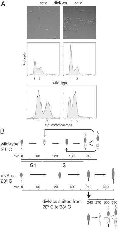
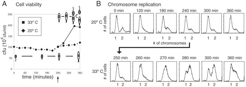
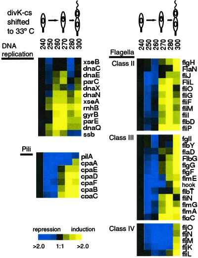
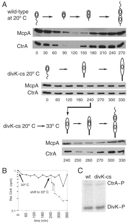
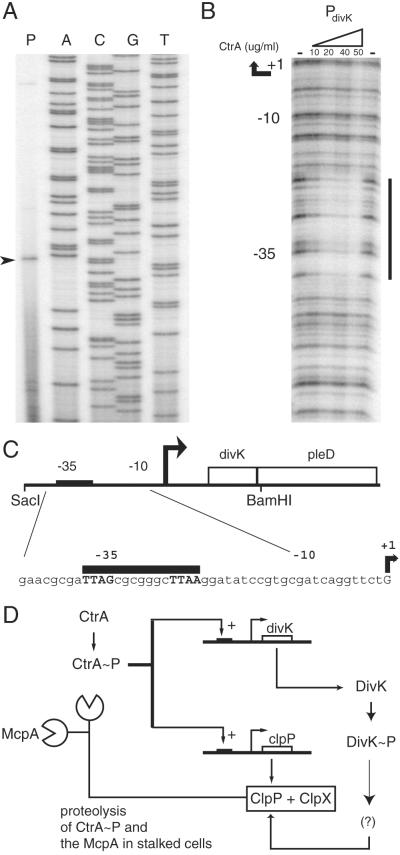
Temporally controlled proteolysis of the essential response regulator, CtrA, is critical for cell cycle progression in Caulobacter crescentus. CtrA binds to and silences the origin of replication in swarmer cells. The initiation of replication depends on the proteolysis of CtrA. We present evidence that DivK, an essential single-domain response regulator, contributes to the control of the G(1)-S transition by signaling the temporally controlled proteolysis of CtrA. In a divK-cs mutant at the restrictive temperature, the initiation of DNA replication is blocked because of the retention of CtrA. A shift of cells from restrictive to permissive temperature results in rapid degradation of CtrA, initiation of DNA replication, and the resumption of cell cycle progression, including the ordered expression of genes involved in chromosome replication and polar organelle biogenesis. CtrA binds to and regulates the promoters of two genes critical to its temporally controlled proteolysis, divK and clpP, providing a transcriptional feedback loop for the control of cell cycle progression.


Figures

Figure 1
Figure 1
A divK-cs mutant at the restrictive temperature blocks the G1–S transition and accumulates elongated stalked cells. (A) Light microscopy of a divK-cs strain grown in rich media at the permissive temperature, 30°C, and at the restrictive temperature, 20°C. Below is the FACS analysis of the divK-cs mutant strain and wild-type C. crescentus. At the restrictive temperature, only the divK-cs cells accumulate single chromosomes. (B) Schematic of the morphology of wild-type and divK-cs synchronous cultures grown at the restrictive temperature (20°C) and divK-cs cells shifted to permissive temperature (33°C) at 240 min. Swarmer cells were isolated from cultures grown at 33°C and then incubated at 20°C in minimal media for 240 min. Wild-type cells differentiated into stalked cells at 90 min, and divided at 270 min, whereas divK-cs stalked cells appeared at 120 min and remained as elongated stalked cells. An aliquot of the divK-cs culture at 20°C shifted back to 33°C at 240 min completed division by 330 min. Shaded areas indicate presence of CtrA.
Figure 2
Figure 2
divK-cs cultures shifted back to permissive temperature complete DNA replication and cell division. (A) At the restrictive temperature, divK-cs cultures had constant colony-forming units (cfu). On shift to the permissive temperature after 240 min at 20°C, the number of colony-forming units doubled, indicating the completion of cell division. (B) FACS analysis of synchronized divK-cs at 20°C and 33°C. At restrictive temperature, cells had predominantly one chromosome. After shift back to permissive temperature at 240 min, cells completed DNA replication by 300 min.
Figure 3
Figure 3
Microarray analysis of total RNA collected at the indicated times after a shift of divK-cs from the restrictive temperature (20°C) back to the permissive temperature (33°C). The expression of genes involved in DNA replication, flagellar biogenesis, and pili biogenesis are shown. RNA was hybridized to microarrays containing 3,700 C. crescentus ORFs. The reference RNA was from a mixed population of cells grown at 33°C. Blue indicates a decrease and yellow indicates an increase relative to the 240-min time point. Black indicates no change. The data represent an average of three experimental repetitions.
Figure 4
Figure 4
(A) Turnover of the CtrA response regulator and the McpA chemoreceptor as a function of the cell cycle in wild-type (wt) and divK-cs cells grown at the restrictive temperature. CtrA is cleared from wild-type cells by 90 min and reappears by 150 min. McpA is also cleared from stalked cells but somewhat later than CtrA. In the synchronized divK-cs mutant at 20°C, both proteins are present through 330 min. After 240 min at 20°C, divK-cs cells shifted to the permissive temperature (33°C) exhibited turnover of both proteins within 10 min. Accumulation of these proteins resumed within 20 min. (B) CtrA protein is stabilized in a divK-cs mutant carrying ctrA under the control of a xylose-inducible promoter (pID42). The pID42/divK-cs strain was grown at 33°C in M2G minimal media and isolated swarmer cells pulsed for 7 min with [32S]Met simultaneous with xylose induction (0.3%). Growth continued at 20°C and at 240 min a portion of the culture was shifted back to 33°C, immunoprecipitated with antibody to CtrA, and separated proteins were analyzed on a Molecular Dynamics PhosphorImager. (C) Wild-type and divK-cs cells at the same OD660 of 0.3 were labeled with [32P]H3PO4, and both DivK and CtrA proteins were immunoprecipitated with their respective antisera. Phosphorylation was visualized on a Molecular Dynamics PhosphorImager. CtrA∼P and DivK∼P are present at comparable levels in both the divK-cs mutant and wild-type.
Figure 5
Figure 5
(A) Primer extension analysis of the divK promoter indicates that the divK mRNA begins at a G residue 55 bp upstream of the protein start site. (B) CtrA∼P footprints the divK promoter at a site overlapping the −35 region. DNaseI protection assays were performed on a PCR-amplified fragment of the divK promoter region. Concentrations of CtrA∼P are indicated above each lane. The minus sign indicates no CtrA protein was added. (C) A schematic of the divK promoter. The CtrA consensus binding site is indicated by a black bar. (D) A model of the regulatory interactions of CtrA, DivK, and the ClpP component of the ClpXP protease. CtrA∼P transcriptionally activates divK and clpP (5, 16). DivK∼P either activates a factor required for ClpXP-mediated proteolysis of CtrA∼P, or inactivates a repressor of ClpXP-mediated proteolysis of CtrA∼P.

References

    1. Quon K C, Marczynski G T, Shapiro L. Cell. 1996;84:83–93. - PubMed
    1. Hecht G B, Lane T, Ohta N, Sommer J M, Newton A. EMBO J. 1995;14:3915–3924. - PMC - PubMed
    1. Jenal U. FEMS Microbiol Rev. 2000;24:177–191. - PubMed
    1. Jacobs C, Domian I J, Maddock J R, Shapiro L. Cell. 1999;97:111–120. - PubMed
    1. Laub M T, Chen S L, Shapiro L, McAdams H H. Proc Natl Acad Sci USA. 2002;99:4632–4637. - PMC - PubMed

Publication types

MeSH terms

LinkOut - more resources