. 2002 Oct;11(10):2285-96.
doi: 10.1110/ps.0218302.

CxxS: fold-independent redox motif revealed by genome-wide searches for thiol/disulfide oxidoreductase function

Affiliations

CxxS: fold-independent redox motif revealed by genome-wide searches for thiol/disulfide oxidoreductase function

Dmitri E Fomenko et al. Protein Sci. 2002 Oct.

Abstract

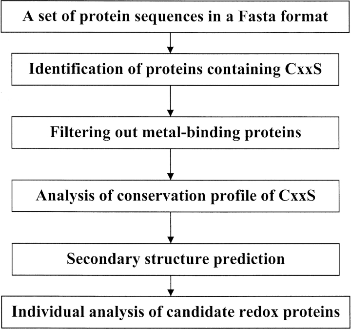
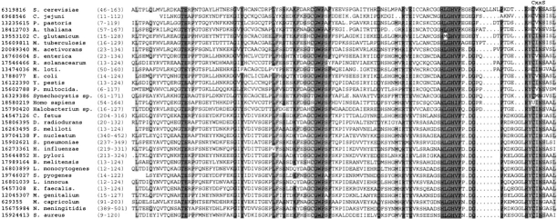
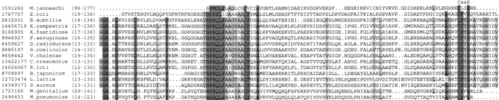
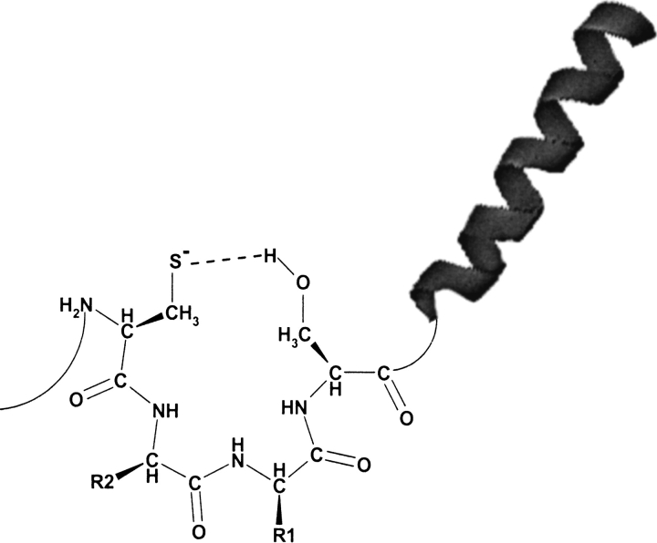
Redox reactions involving thiol groups in proteins are major participants in cellular redox regulation and antioxidant defense. Although mechanistically similar, thiol-dependent redox processes are catalyzed by structurally distinct families of enzymes, which are difficult to identify by available protein function prediction programs. Herein, we identified a functional motif, CxxS (cysteine separated from serine by two other residues), that was often conserved in redox enzymes, but rarely in other proteins. Analyses of complete Escherichia coli, Campylobacter jejuni, Methanococcus jannaschii, and Saccharomyces cerevisiae genomes revealed a high proportion of proteins known to use the CxxS motif for redox function. This allowed us to make predictions in regard to redox function and identity of redox groups for several proteins whose function previously was not known. Many proteins containing the CxxS motif had a thioredoxin fold, but other structural folds were also present, and CxxS was often located in these proteins upstream of an alpha-helix. Thus, a conserved CxxS sequence followed by an alpha-helix is typically indicative of a redox function and corresponds to thiol-dependent redox sites in proteins. The data also indicate a general approach of genome-wide identification of redox proteins by searching for simple conserved motifs within secondary structure patterns.


Figures

Fig. 1.
Fig. 1.
Schematic representation of the algorithm for identification of thiol/disulfide oxidoreductases containing the CxxS motif that was used in our study.
Fig. 2.
Fig. 2.
Alignment of methionine-R-sulfoxide reductases. The location of the CxxS motif in C-terminal regions of the enzymes is indicated above the sequences. Residues conserved in >90% of the sequences are highlighted with dark gray. Residues conserved in >70% of the sequences are highlighted with light gray. Accession numbers (gi) for each sequence used in the alignment are indicated on the left. In vertebrate methionine-R-sulfoxide reductase, selenoprotein R, the cysteine in the CxxS motif is replaced with selenocysteine (shown as U for human enzyme).
Fig. 3.
Fig. 3.
Alignment of proteins of the OsmC family. The location of the CxxS motif in C-terminal regions of the protein is indicated above the sequences. Residues conserved in >90% of the sequences are highlighted with dark gray. Residues conserved in >60% of the sequences are highlighted with light gray. Accession numbers (gi) for each sequence used in the alignment are indicated on the left.
Fig. 4.
Fig. 4.
Alignment of proteins of the Yhe family. The location of the CxxS motif in C-terminal regions of the proteins is indicated above the sequences. Residues conserved in >80% of the sequences are highlighted with dark gray. Residues conserved in >60% of the sequences are highlighted with light gray. Accession numbers (gi) for each sequence used in the alignment are indicated on the left.
Fig. 5.
Fig. 5.
Alignment of DsrE-like proteins. The location of the CxxS motif in C-terminal regions of the proteins is indicated above the sequences. Residues conserved in 100% of the sequences are highlighted with dark gray. Residues conserved in >60% of the sequences are highlighted with light gray. Accession numbers (gi) for each sequence used in the alignment are indicated on the left.
Fig. 6.
Fig. 6.
A model for the involvement of serine and the α-helix in stabilization of cysteine thiolate involved in redox reactions. The presence of an α-helix downstream of the CxxS sequence could also be owing to frequent occurrence of these structures near catalytic surface-exposed loops.

Similar articles

Cited by

References

    1. Altschul, S.F., Madden, T.L., Schaffer, A.A., Zhang, J., Zhang, Z., Miller, W., and Lipman, D. 1997. Gapped BLAST and PSI-BLAST: A new generation of protein database search programs. Nucleic Acids Res. 25 3389–3402. - PMC - PubMed
    1. Anelli, T., Alessio, M., Mezghrani, A., Simmen, T., Talamo, F., Bachi, A., and Sitia, R. 2002. ERp44, a novel endoplasmic reticulum folding assistant of the thioredoxin family. EMBO J. 21 835–844. - PMC - PubMed
    1. Arner, E.S. and Holmgren, A. 2000. Physiological functions of thioredoxin and thioredoxin reductase. Eur. J. Biochem. 267 6102–6109. - PubMed
    1. Atichartpongkul, S., Loprasert, S., Vattanaviboon, P., Whangsuk, W., Helmann, J.D., and Mongkolsuk, S. 2001. Bacterial Ohr and OsmC paralogues define two protein families with distinct functions and patterns of expression. Microbiology 147 1775–1782. - PubMed
    1. Bateman, A. 1999. The SIS domain: A phosphosugar-binding domain. Trends. Biochem. Sci. 24 94–95. - PubMed

MeSH terms

Substances

LinkOut - more resources