. 2002 Oct;11(10):2335-50.
doi: 10.1110/ps.4270102.

Molecular dynamics simulations of alanine rich beta-sheet oligomers: Insight into amyloid formation

Affiliations

Molecular dynamics simulations of alanine rich beta-sheet oligomers: Insight into amyloid formation

Buyong Ma et al. Protein Sci. 2002 Oct.

Abstract

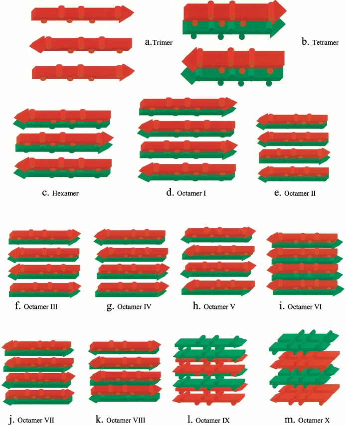
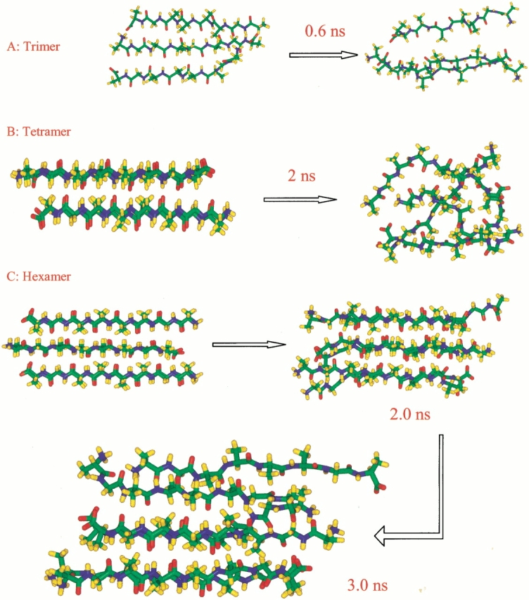
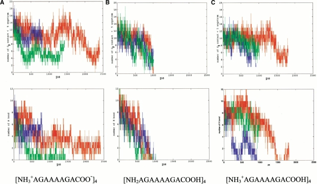
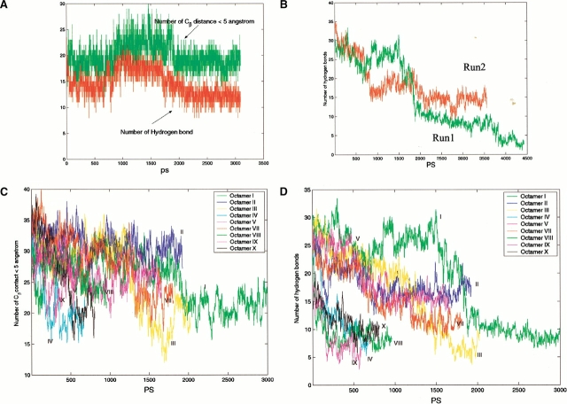
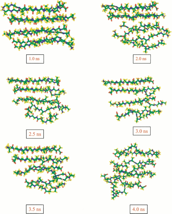
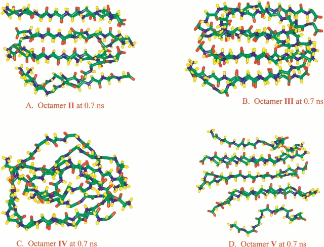
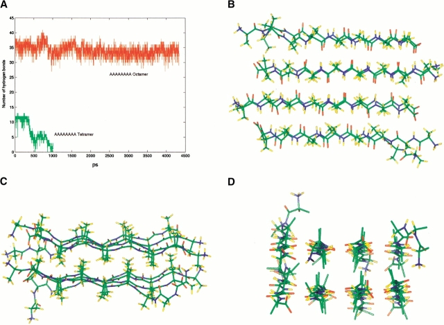
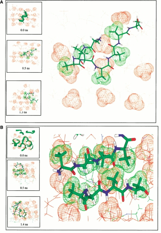
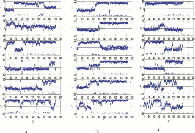
The aggregation observed in protein conformational diseases is the outcome of significant new beta-sheet structure not present in the native state. Peptide model systems have been useful in studies of fibril aggregate formation. Experimentally, it was found that a short peptide AGAAAAGA is one of the most highly amyloidogenic peptides. This peptide corresponds to the Syrian hamster prion protein (ShPrP) residues 113-120. The peptide was observed to be conserved in all species for which the PrP sequence has been determined. We have simulated the stabilities of oligomeric AGAAAAGA and AAAAAAAA (A8) by molecular dynamic simulations. Oligomers of both AGAAAAGA and AAAAAAAA were found to be stable when the size is 6 to 8 (hexamer to octamer). Subsequent simulation of an additional alpha-helical AAAAAAAA placed on the A8-octamer surface has revealed molecular events related to conformational change and oligomer growth. Our study addresses both the minimal oligomeric size of an aggregate seed and the mechanism of seed growth. Our simulations of the prion-derived 8-residue amyloidogenic peptide and its variant have indicated that an octamer is stable enough to be a seed and that the driving force for stabilization is the hydrophobic effect.


Figures

Fig. 1.
Fig. 1.
Oligomer conformations investigated in this work. Peptides in the same sheet have the same color. The sequence is AGAAAAGA, with the alanine methyl groups depicted as cylindrical humps. (a) Trimer in an antiparallel sheet. (b) Tetramer in two layers. In each layer, the strands are antiparallel. The sheets are parallel. (c) Hexamer in two layers. In each layer the strands are antiparallel sheet. The sheets are parallel. (d) Octamer I. The most stable octamer orientation in which the β strands are antiparallel within each sheet layer but parallel across layers. (e) Octamer II. Three β-strands are antiparallel and one strand is parallel with parallel layers. (f) Octamer III. One parallel interaction (the two strands in the center) and two anti-parallel interactions (at the edges of the complex) with parallel stacking across layers. (g) Octamer IV. Two parallel interactions (along the edges of the complex) and one antiparallel interaction (in the center of the complex) with parallel layers. (h) Octamer V. The β-strands are antiparallel both within each β-sheet layer and across layers. (i) Octamer VI. The β-strands are parallel within each β-sheet layer and with antiparallel layers. (j) Octamer VII. Two parallel interactions (along the edges of the complex) and one antiparallel interaction with antiparallel layers. (k) Octamer VIII. One parallel interaction (two strands in the center) and two antiparallel interactions (at the edges of the complex), with antiparallel stacking across layers. (l) Octamer IX. A sheet of two antiparallel β-strands stacks on other two-stranded sheets in four parallel layers. (m) Octamer X. A sheet of two parallel β-strands stacks on other two-stranded sheets in four antiparallel layers.
Fig. 2.
Fig. 2.
Snapshots of the conformational changes of trimer (a), tetramer (b), and hexamer (c). The initial conformations are those depicted schematically in Figure 1a–c ▶, respectively.
Fig. 3.
Fig. 3.
Trajectories (methyl group contacts, top row; hydrogen bonds, bottom row) for the tetramer in three ionic states. (a) zwitterion, (b) neutral, and (c) positively charged. Three simulations are performed for each ionic state.
Fig. 4.
Fig. 4.
Trajectories from simulations of the following: (a) Hexamer, showing the Cβ contacts and the number of hydrogen bonds. (b) Two trajectories of Octamer I in the most stable orientation (Fig. 1d ▶). The figure shows the number of interstrand hydrogen bonds from the two simulations. (c) The methyl group contacts from the simulation of all octamers (Fig. 1e–m ▶), except for Octamer VI. This octamer dissociated after 300 ps and thus is not shown here. (d) The number of interstrand hydrogen bonds from simulations of all octamers, except for Octamer VI. The schematic initial conformations are given in Figure 1e–m ▶.
Fig. 5.
Fig. 5.
Snapshots of conformational changes taking place in Octamer I (Fig. 1d ▶) from run 1. The stable β-strand interactions are retained over a long simulation time.
Fig. 6.
Fig. 6.
Snapshots from the simulations for several octamers at 0.7 nanoseconds (ns) simulation time. (a) Octamer II. (b) Octamer III. (c) Octamer IV. (d) Octamer V. The initial conformations are schematically drawn in Figure 1 ▶.
Fig. 7.
Fig. 7.
Polyalanine simulations: (a) Trajectories of the (NH3+AAAAAAAACOO)8 octamer and of the (NH3+AAAAAAAACOO)4 tetramer. (b–d) Different views of a snapshot from the octamer polyalanine simulations.
Fig. 8.
Fig. 8.
The conformational change of additional octa-alanines (green color) placed on the polyalanine octamer surface (red). Only methyl groups on the top of the surface are shown for the octamer. The large figures are enhanced views of the atomic details of the surface matching. The inserted boxes are snapshots from the simulations. (a) Simulation of a preformed octamer plus an α-helical octa-alanine at 350 K. The green methyl groups of (A3A4A5A6A7) in the additional octa-alanine indicate the match with the methyl groups on the octamer surface (in red). (b) Simulation of a preformed octamer plus eight random octa-alanines at 400 K. The enlarged figure shows the perfect match of the β-strands patches on the lateral surface, which lasts about 0.25 ns. No β-interactions are observed between the octamer and the random peptides along the edge.
Fig. 8.
Fig. 8.
The conformational change of additional octa-alanines (green color) placed on the polyalanine octamer surface (red). Only methyl groups on the top of the surface are shown for the octamer. The large figures are enhanced views of the atomic details of the surface matching. The inserted boxes are snapshots from the simulations. (a) Simulation of a preformed octamer plus an α-helical octa-alanine at 350 K. The green methyl groups of (A3A4A5A6A7) in the additional octa-alanine indicate the match with the methyl groups on the octamer surface (in red). (b) Simulation of a preformed octamer plus eight random octa-alanines at 400 K. The enlarged figure shows the perfect match of the β-strands patches on the lateral surface, which lasts about 0.25 ns. No β-interactions are observed between the octamer and the random peptides along the edge.
Fig. 9.
Fig. 9.
Comparison of ξ angle changes of polyalanine in isolated state (a) and on the octamer surface (b, 350 K simulation; c, 400 K simulation). The angles are ξ1: N2-Cα2-C2-N3; ξ2: N3-Cα3-C3-N4; ξ3: N4-Cα4-C4-N5; ξ4: N5-Cα5-C5-N6; ξ5: N6-Cα6-C6-N7; |gR6: N7-Cα7-C7-N8.

Similar articles

Cited by

References

    1. Azriel, R. and Gazit, E. 2001. Analysis of the minimal amyloid-forming fragment of the islet amyloid polypeptide. J. Biol. Chem. 276 34156–34161. - PubMed
    1. Balbach, J.J., Ishii, Y., Antzutkin, O.N., Leapman, R.D., Rizzo, N.W., Dyda, F., Reed, J., and Tycko, R. 2000. Amyloid fibril formation by Aβ16–22, a seven-residue fragment of the Alzheimer's β-amyloid peptide, and structural characterization by solid state NMR. Biochemistry 39 13748–13759. - PubMed
    1. Balbirnie, M., Grothe, R., and Eisenberg, D.S. 2001. An amyloid-forming peptide from the yeast prion Sup35 reveals a dehydrated β-sheet structure from amyloid. Proc. Natl. Acad. Sci. 98 2375–2380. - PMC - PubMed
    1. Benziger, T.L.S., Gregory, D.M. Burkoth, T.S., Miller-Auer, H., Lynn, D.G., Botto, R.E., and Meredith, S.C. 2000. Two-dimensional structure of β-amyloid(10–35) fibrils. Biochemistry 39 3491–3499. - PubMed
    1. Blondelle, S.B., Forood, B., Houghten, R.A., and Perez-Paya, E. 1996. Polyalanine-based peptides as models for self-associated β-pleated-sheet complexes. Biochemistry 36 8393–8400. - PubMed

Publication types