

Akt induces enhanced myocardial contractility and cell size in vivo in transgenic mice
- PMID: 12237475
- PMCID: PMC129445
- DOI: 10.1073/pnas.172376399
Akt induces enhanced myocardial contractility and cell size in vivo in transgenic mice
Abstract
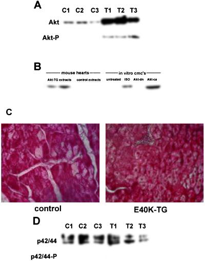
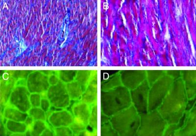
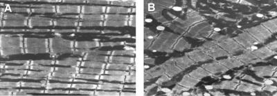
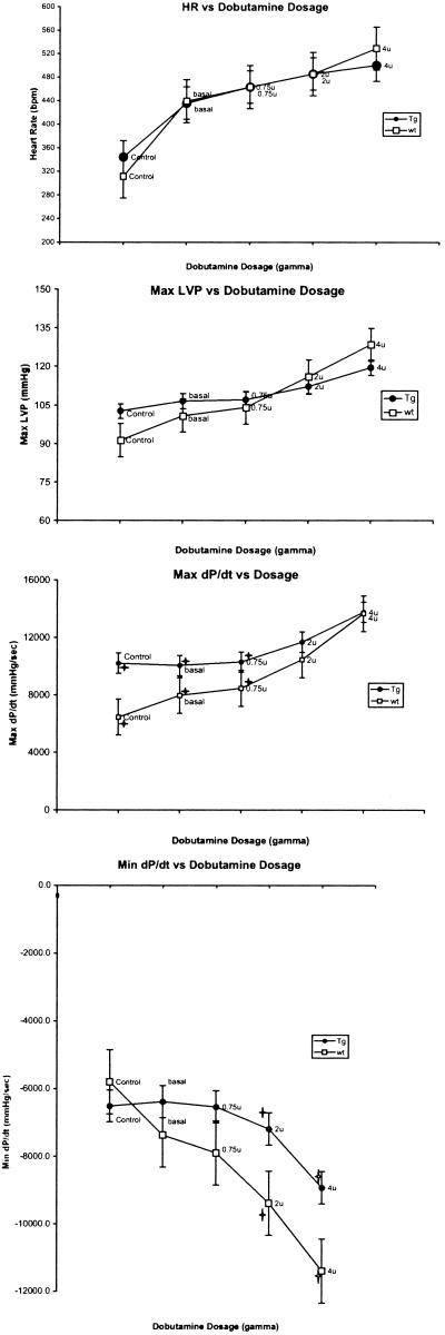
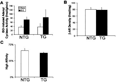
The serine-threonine kinase Akt seems to be central in mediating stimuli from different classes of receptors. In fact, both IGF-1 and IL6-like cytokines induce hypertrophic and antiapoptotic signals in cardiomyocytes through PI3K-dependent Akt activation. More recently, it was shown that Akt is involved also in the hypertrophic and antiapoptotic effects of beta-adrenergic stimulation. Thus, to determine the effects of Akt on cardiac function in vivo, we generated a model of cardiac-specific Akt overexpression in mice. Transgenic mice were generated by using the E40K, constitutively active mutant of Akt linked to the rat alpha-myosin heavy chain promoter. The effects of cardiac-selective Akt overexpression were studied by echocardiography, cardiac catheterization, histological and biochemical techniques. We found that Akt overexpression produced cardiac hypertrophy at the molecular and histological levels, with a significant increase in cardiomyocyte cell size and concentric LV hypertrophy. Akt-transgenic mice also showed a remarkable increase in cardiac contractility compared with wild-type controls as demonstrated by the analysis of left ventricular (dP/dt(max)) in an invasive hemodynamic study, although with graded dobutamine infusion, the maximum response was not different from that in controls. Diastolic function, evaluated by left ventricular dP/dt(min), was not affected at rest but was impaired during graded dobutamine infusion. Isoproterenol-induced cAMP levels, beta-adrenergic receptor (beta-AR) density, and beta-AR affinity were not altered compared with control mice. Moreover, studies on signaling pathway activation from myocardial extracts demonstrated that glycogen synthase kinase3-beta is phosphorylated, whereas p42/44 mitogen-activated protein kinases is not, indicating that Akt induces hypertrophy in vivo by activating the glycogen synthase kinase3-beta/GATA 4 pathway. In summary, our results not only demonstrate that Akt regulates cardiomyocyte cell size in vivo, but, importantly, show that Akt modulates cardiac contractility in vivo without directly affecting beta-AR signaling capacity.
Figures
References
-
- Bellacosa A., Chan, T. O., Ahmed, N. N., Datta, K., Malstrom, S., Stokoe, D., McCormick, F., Feng, J. & Tsichlis, P. (1998) Oncogene 17, 313-325. - PubMed
-
- Oh H., Fujio, Y., Kunisada, K., Hirota, H., Matsui, H., Kishimoto, T. & Yamauchi-Takihara, K. (1998) J. Biol. Chem. 273, 9703-9710. - PubMed
-
- Morisco C., Zebrowski, D., Condorelli, G., Tsichlis, P., Vatner, S. F. & Sadoshima, J. (2000) J. Biol. Chem. 275, 14466-14475. - PubMed
Publication types
MeSH terms
Substances
LinkOut - more resources
Full Text Sources
Other Literature Sources
Molecular Biology Databases
Research Materials
Miscellaneous
