Comparative Study
. 2002 Sep 9;87(6):635-44.
doi: 10.1038/sj.bjc.6600510.

Rho GTPases in human breast tumours: expression and mutation analyses and correlation with clinical parameters

Affiliations
Comparative Study

Rho GTPases in human breast tumours: expression and mutation analyses and correlation with clinical parameters

G Fritz et al. Br J Cancer. .

Abstract

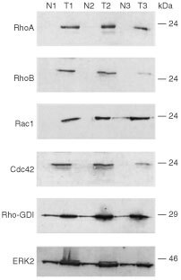
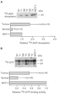
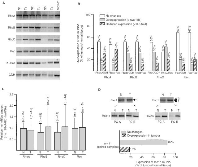
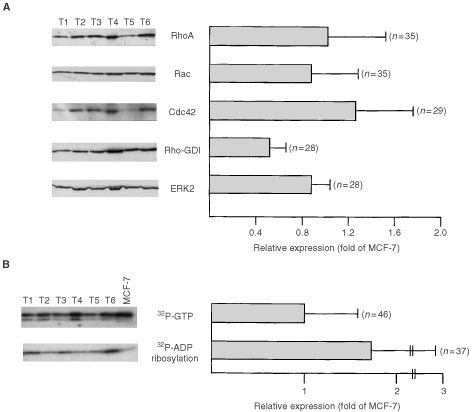
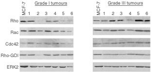
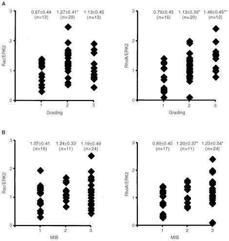
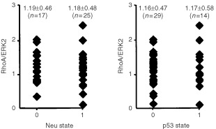
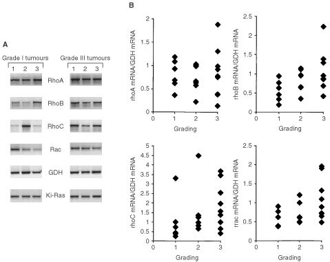
In the present study, we addressed the question of a putative relevance of Rho proteins in tumour progression by analysing their expression on protein and mRNA level in breast tumours. We show that the level of RhoA, RhoB, Rac1 and Cdc42 protein is largely enhanced in all tumour samples analysed (n=15) as compared to normal tissues originating from the same individual. The same is true for (32)P-ADP-ribosylation of Rho proteins which is catalysed by Clostridium botulinum exoenzyme C3. Also the amount of Rho-GDI and ERK2 as well as the level of overall (32)P-GTP binding activity was tumour-specific elevated, yet to a lower extent than Rho proteins. Although the amount of Rho proteins was enhanced in tumours, most of them did not show changes in rho mRNA expression as compared to the corresponding normal tissue. Thus, elevated gene expression seems not to be the underlying mechanism of tumour-specific overexpression of Rho proteins. Sequence analysis of RhoA, RhoB, RhoC and Rac1 failed to detect any mutations in both the GTP-binding site and effector binding region. By analysing >50 tumour samples, the amount of RhoA-like proteins (i.e. RhoA, B, C), but not of Rac1, was found to significantly increase with histological grade and proliferation index. Rho protein expression was neither related to p53 nor to HER-2/neu oncogene status. Expression of rho mRNAs did not show a significant increase with histological grade. Overall the data show that (1) Rho proteins are overexpressed in breast tumours (2) overexpression is not regulated on the mRNA level (3) the expression level of RhoA-like proteins correlates with malignancy and (4) Rho proteins are not altered by mutation in breast tumours.


Figures

Figure 1
Figure 1
Expression of Rho GTPases in malignant and non-malignant breast tissue. Each 50 μg of protein isolated from tumorigenic (T1, T2, T3) and corresponding normal breast tissue (N1, N2, N3) originating from the same patient was separated by SDS–PAGE and subjected to Western blot analysis using the antibodies indicated. Shown is the autoradiography.
Figure 2
Figure 2
Comparative analysis of C3-mediated 32P-ADP-ribosylation and 32P-GTP binding activity of Rho proteins in human breast tumours as compared to normal tissue. (A) 32P-ADP-ribosylation of Rho proteins in extracts from tumour and corresponding non-malignant tissue. Twenty-five μg of cytosolic protein was 32P-ADP-ribosylated by use of exoenzyme C3 from C. botulinum as described in Materials and Methods. Reaction products were separated by SDS–PAGE. Autoradiography was densitometrically analysed and relative 32P-ADP-ribosylation of tissue extracts was related to that of MCF-7 cells, which was set to 1.0. Autoradiogram of a representative analysis of three sampled pairs is shown in the upper part of the figure. N, normal tissue; T, tumour. (B) Analysis of 32P-GTP binding activity of extracts from non-malignant and tumorigenic breast tissue. Thirty μg of protein was separated by SDS–PAGE (15% gels). After wet blotting to nitrocellulose and renaturation, 32P-GTP binding activity was analysed as described in Materials and Methods. Relative GTP binding activity of extracts from MCF-7 cells was set to 1.0. The autoradiogram of a representative analysis is shown in the upper part of the figure. N, normal tissue; T, tumour.
Figure 3
Figure 3
rho mRNA expression in tumorigenic and normal breast tissue. (A) Total RNA from tumorigenic and corresponding normal tissue from breast was analysed as to the expression of various rho mRNA species (i.e. rhoA, rhoB, rhoC, rac1) by RT–PCR analysis as described in Materials and Methods. As internal control expression of Ki-ras and GDH mRNA was determined. N, normal tissue; T, corresponding tumour. (B) Quantitative densitometrical analysis of relative rho mRNA expression in breast tumours vs normal tissue. Shown is the percentage of tumours showing similar, increased or reduced level of rho mRNA expression as compared to the corresponding normal tissue of the same individual. Relative expression of rho mRNA species was calculated by referring rho mRNA amount to that of either Ki-Ras (Ras) or GDH (GDH). Both types of internal reference markers gave the same results. (C) Interindividual variation in the relative expression of rho mRNAs (rho mRNA/GDH mRNA) in normal and tumorigenic tissues from breast. (D) The upper part of the figure shows the results of representative RT–PCR analyses of two different paired samples (normal tissue (N) vs tumorigenic tissue (T)) where rac1b is clearly detectable (right part) or not (left part) as a co-amplification product of the rac1 RT–PCR reaction (Rac1). 1,2, indicates the position of rac1 and rac1b amplification product, respectively. For more specific and sensitive analysis of rac1b expression (Rac1b), nested PCR was performed using two different types of rac1b specific primer combinations as described in Materials and Methods (PC-A and PC-B). In the lower part of the figure the quantitative evaluation of the expression of rac1b in normal breast tissue vs the corresponding tumorigenic tissue from the same patient (n=11; paired samples) is shown. Please note that the samples representatively shown under A and D are different from each other.
Figure 4
Figure 4
Analysis of expression of Rho proteins, ADP-ribosylated proteins and GTP-binding proteins in breast tumour samples. (A) Expression of Rho proteins (RhoA-like proteins (Rho), Rac1 and Cdc42) and Rho-regulatory factor Rho-GDI in breast tumours was analysed by Western blot analysis. Additionally the amount of ERK2 protein was determined. For statistical analysis, expression of Rho and Rho-GDI proteins in tumours was related to that of MCF-7 cells which was set to 1.0. In the left part of the figure an autoradiogram of a representative analysis of six tumour samples is shown. Rho, the Rho antibody used cross-reacts with the RhoA-like Rho GTPases RhoA, B and C. (B) Variability in 32P-GTP-binding activity and 32P-ADP-ribosylation of extracts from breast tumours. For statistical analysis, GTP-binding activity and level of 32P-ADP-ribosylation of breast tissue extracts was related to that of MCF-7 cells which was set to 1.0. In the left part of the figure, an autoradiography of a representative analysis of six tumour samples is shown.
Figure 5
Figure 5
Comparative expression of Rho GTPases in breast tumours of different histological grade. Six representative samples of human breast tumours of grade I and grade III, respectively, were analysed by Western blot analysis as to the expression of various Rho GTPase, the Rho-regulatory factor Rho-GDI and ERK2. As a control, extracts from MCF-7 cells were included.
Figure 6
Figure 6
Rho protein expression correlates with histological grade and proliferation index of breast tumours. (A) Relative expression of RhoA-like and Rac1 proteins of breast tumours was calculated by referring the Rho protein amount to the level of ERK2 protein (RhoA/ERK2 and Rac/ERK2, respectively). Relative Rho protein expression was related to the histological grade of breast tumours. 1, 2 and 3; grade I, grade II and grade III.*, statistical significance as referred to grade I (*P<0.05; **P<0.001). (B) Relative RhoA and Rac1 protein expression (RhoA/ERK2; Rac/ERK2) of breast tumours was related to proliferation index (MIB-1) of the tumours. 1, 2, 3: <5%, <20%, >20% MIB-1 positive cells.*, statistical significance as referred to MIB-1 <5% (*P<0.05).
Figure 7
Figure 7
Rho protein expression is not related to HER-2/neu oncogene or p53 status of breast tumours. Relative expression of RhoA-like Rho GTPases was related to HER-2/neu oncogene and p53 status of the tumours. 0, negative for either HER-2/neu overexpression or p53 mutation; 1, positive for HER-2/neu overexpression or p53 mutation.
Figure 8
Figure 8
Relationship between rho mRNA expression and histological grade of breast tumours. (A) Representative analysis of rho mRNA expression (i.e. rhoA, rhoB, rhoC, rac1) in breast tumours of WHO grade I and III, respectively. As internal control mRNA expression of GDH and Ki-Ras was determined. (B) Correlation analysis of rho mRNA expression and grading status of breast tumours. Relative rho mRNA expression was determined by referring rho mRNA levels to that of GDH mRNA. 1,2,3; grade I, II and III, respectively.

References

    1. AktoriesK1997Bacterial toxins that target Rho proteins J Clin Invest 99827829 - PMC - PubMed
    1. AmanoMItoMKimuraKFukutaYChiharaKNakanoTMatsuuraYKaibuchiK1996Phosphorylation and activation of myosin by Rho-associated kinase (Rho-kinase) J Biol Chem 2712024620249 - PubMed
    1. AndersonMWReynoldsSHYouMMaronpotRM1992Role of Proto-oncogene activation in carcinogenesis Environm. Health Perspectives 981324 - PMC - PubMed
    1. AvrahamHWeinbergRA1989Characterization and expression of the human rhoH12 gene product Mol Cell Biol 920582066 - PMC - PubMed
    1. BosJL1988The ras gene family and human carcinogenesis Mut Res 195255271 - PubMed

Publication types

MeSH terms