. 2002 Oct;76(20):10109-21.
doi: 10.1128/jvi.76.20.10109-10121.2002.

Naturally occurring substitutions in the P/V gene convert the noncytopathic paramyxovirus simian virus 5 into a virus that induces alpha/beta interferon synthesis and cell death

Affiliations

Naturally occurring substitutions in the P/V gene convert the noncytopathic paramyxovirus simian virus 5 into a virus that induces alpha/beta interferon synthesis and cell death

Elizabeth K Wansley et al. J Virol. 2002 Oct.

Abstract

The V protein of the paramyxovirus simian virus 5 (SV5) is responsible for targeted degradation of STAT1 and the block in alpha/beta interferon (IFN-alpha/beta) signaling that occurs after SV5 infection of human cells. We have analyzed the growth properties of a recombinant SV5 that was engineered to be defective in targeting STAT1 degradation. A recombinant SV5 (rSV5-P/V-CPI-) was engineered to contain six naturally occurring P/V protein mutations, three of which have been shown in previous transfection experiments to disrupt the V-mediated block in IFN-alpha/beta signaling. In contrast to wild-type (WT) SV5, human cells infected with rSV5-P/V-CPI- had STAT1 levels similar to those in mock-infected cells. Cells infected with rSV5-P/V-CPI- were found to express higher-than-WT levels of viral proteins and mRNA, suggesting that the P/V mutations had disrupted the regulation of viral RNA synthesis. Despite the inability to target STAT1 for degradation, single-step growth assays showed that the rSV5-P/V-CPI- mutant virus grew better than WT SV5 in all cell lines tested. Unexpectedly, cells infected with rSV5-P/V-CPI- but not WT SV5 showed an activation of a reporter gene that was under control of the IFN-beta promoter. The secretion of IFN from cells infected with rSV5-P/V-CPI- but not WT SV5 was confirmed by a bioassay for IFN. The rSV5-P/V-CPI- mutant grew to higher titers than did WT rSV5 at early times in multistep growth assays. However, rSV5-P/V-CPI- growth quickly reached a final plateau while WT rSV5 continued to grow and produced a final titer higher than that of rSV5-P/V-CPI- by late times postinfection. In contrast to WT rSV5, infection of a variety of cell lines with rSV5-P/V-CPI- induced cell death pathways with characteristics of apoptosis. Our results confirm a role for the SV5 V protein in blocking IFN signaling but also suggest new roles for the P/V gene products in controlling viral gene expression, the induction of IFN-alpha/beta synthesis, and virus-induced apoptosis.


Figures

FIG. 1.
FIG. 1.
rSV5 genome structure and amino acid differences between the P/V region of the CPI+ and CPI− strains of SV5. (A) Structure of rSV5-GFP. The SV5 genome is depicted as a rectangle with vertical bars denoting the intergenic regions. The GFP open reading frame is found between the HN and L genes (19). The location of the StuI-ClaI restriction fragment used to introduce the CPI− P/V mutations is indicated. le, leader; tr, trailer. (B) Amino acid differences in the first 165 residues of the amino-terminal region of the P/V proteins are shown for WT, CPI+, and CPI− strains of SV5. Asterisks denote amino acid differences between CPI+ and CPI− (7, 45).
FIG. 2.
FIG. 2.
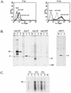
Elevated levels of viral protein and RNA in cells infected with rSV5-V-CPI−. (A) Elevated GFP expression in cells infected with rSV5-V-CPI−. A549 cells were mock infected or infected with rSV5-GFP or rSV5-V-CPI−. The cells were harvested at the indicated times p.i.and analyzed by flow cytometry for expression of GFP. (B) Elevated rate of protein synthesis in cells infected with rSV5-V-CPI−. MDBK cells were mock infected (M lanes) or infected with rSV5-GFP (S lanes) or rSV5-P/V-CPI− (C lanes) at an MOI of 50. At 8 h p.i., the cells were radiolabeled for 20 min using Tran 35S-label, and proteins were immunoprecipitated from cells extracts by using antibodies to NP, P, M, GFP, or V and then analyzed by SDS-PAGE. (C) Elevated mRNA accumulation in cells infected with rSV5-V-CPI−. MDBK cells were mock infected (M lane) or infected with rSV5-GFP (S lanes) or rSV5-P/V-CPI− (C lanes) at an MOI of 50. At the indicated times p.i., poly(A)+ RNA was isolated and analyzed by Northern blotting with a negative-sense NP-specific riboprobe.
FIG. 3.
FIG. 3.
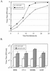
rSV5-P/V-CPI− grows to higher titers than rSV5-GFP. (A) Single-step growth assay. A549 cells were infected at an MOI of 50 with rSV5-GFP or rSV5-P/V-CPI−, and virus titers were determined at the indicated times p.i. by a plaque assay. The results shown are from one of three independent experiments. (B) Virus yield in different cell types. The indicated cell lines were infected at an MOI of 50 with rSV5-GFP or rSV5-P/V-CPI−. At 16 h p.i., medium was harvested and the virus titer was determined by a plaque assay. Data are the average of three determinations, and the standard deviation is shown by bars.
FIG. 4.
FIG. 4.
Protein expression and STAT1 levels in cells infected with rSV5-GFP or rSV5-P/V-CPI− virus. (A) Infection of human cells with rSV5-P/V-CPI− does not induce STAT1 degradation. A549 cells were mock infected (lanes 1) or infected with rSV5-P/V-CPI− (lanes 2) or rSV5-GFP (lanes 3) at an MOI of ∼50. At 24 h p.i., the cells were lysed and equivalent amounts of protein were analyzed by Western blotting with antisera specific for STAT1 (left) or SV5 P protein (right). (B) Time course of P-protein and STAT1 levels. A549 cells were infected with rSV5-GFP (left) or rSV5-P/V-CPI− (right) at an MOI of 50. At the indicated times p.i., cells were lysed and equivalent amounts of protein were analyzed by Western blotting with antisera specific for the SV5 P protein (top) or STAT1 (bottom).
FIG. 5.
FIG. 5.
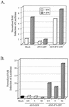
IFN signaling in cells infected with rSV5-V-CPI−. (A) Induction of ISRE. A549 cells were cotransfected with pSV-βgal and a plasmid containing the luciferase gene under the control of an ISRE. At 24 h later, the cells were mock infected or infected with rSV5-GFP or rSV5-P/V-CPI− at an MOI of 50. At 16 h p.i., the cells were incubated for 6 h with (hatched bars) or without (white bars) 2,000 U of IFN-α/β. Normalized luciferase activity was calculated by dividing luciferase activity by β-gal activity and is expressed as a fold induction over that seen in mock-infected cells. Data are representative of three independent experiments. (B) Effect of virus dose on the induction of ISRE. A549 cells were treated as described for panel A, except that the infections were at an MOI of 0.5, 5, or 50. At 20 h postinfection, normalized luciferase activity was determined as described for panel A. Data are representative of three independent experiments.
FIG. 6.
FIG. 6.
Induction of the IFN-β promoter by infection with rSV5-V-CPI− but not WT rSV5-GFP. A549 cells were transiently cotransfected with pSV-βgal and a plasmid encoding the luciferase gene under the control of the IFN-β promoter. At 24 h p.i., the cells were mock infected or infected with rSV5-GFP or rSV5-P/V-CPI− at the indicated MOI. Cells were lysed at 24 h p.i., and normalized luciferase activity was calculated as a fold induction over that seen in mock-infected cells. Data are the average of three determinations, and the standard deviation is shown by bars.
FIG. 7.
FIG. 7.
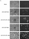
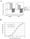
Multiple-step growth with WT rSV5-GFP and rSV5-P/V-CPI−. (A) Fluorescence microscopy assay for virus spread. A549 cells were mock infected or infected at an MOI of 0.05 with rSV5-GFP or rSV5-P/V-CPI−. At 24 or 48 h p.i., the cells were fixed with 2% PFA and then analyzed by microscopy for GFP fluorescence. Phase-contrast microscopy is shown in the left panels, and GFP fluorescence is shown in the right panels. (B) Multiple-step growth assay. A549 cells were infected with rSV5-GFP or rSV5-P/V-CPI− at an MOI of 0.05. Medium was harvested at the indicated times postinfection, and the viral titer was determined by a plaque assay on CV-1 cells.
FIG. 7.
FIG. 7.
Multiple-step growth with WT rSV5-GFP and rSV5-P/V-CPI−. (A) Fluorescence microscopy assay for virus spread. A549 cells were mock infected or infected at an MOI of 0.05 with rSV5-GFP or rSV5-P/V-CPI−. At 24 or 48 h p.i., the cells were fixed with 2% PFA and then analyzed by microscopy for GFP fluorescence. Phase-contrast microscopy is shown in the left panels, and GFP fluorescence is shown in the right panels. (B) Multiple-step growth assay. A549 cells were infected with rSV5-GFP or rSV5-P/V-CPI− at an MOI of 0.05. Medium was harvested at the indicated times postinfection, and the viral titer was determined by a plaque assay on CV-1 cells.
FIG. 8.
FIG. 8.
Infection with rSV5-P/V-CPI− but not WT rSV5-GFP results in cell death. (A) Cell viability during infection with rSV5-GFP or rSV5-V-CPI−. MDBK or A549 cells were mock infected or infected with rSV5-GFP or rSV5-P/V-CPI− at an MOI of 50. At 2 days p.i., viable-cell numbers were determined by trypan blue staining. Data are expressed as a fold change in cell number from the starting culture. (B) Time-lapse video microscopy of cytopathic effect. A549 cells were infected with rSV5-GFP or rSV5-P/V-CPI− at an MOI of ∼50, and cells were monitored by time-lapse video microscopy. At the indicated times p.i., the number of rounded cells in the field that did not later divide was counted as a percentage of the total number of cells in the field. Data are the mean of three determinations, and the standard deviation is shown by bars. (C) Fluorescence microscopy of TUNEL staining for DNA fragmentation. A549 cells were mock infected or infected at an MOI of 50. At 48 h p.i., TUNEL staining was performed as described in Materials and Methods. Photographs were taken at the same exposure for all samples.
FIG. 8.
FIG. 8.
Infection with rSV5-P/V-CPI− but not WT rSV5-GFP results in cell death. (A) Cell viability during infection with rSV5-GFP or rSV5-V-CPI−. MDBK or A549 cells were mock infected or infected with rSV5-GFP or rSV5-P/V-CPI− at an MOI of 50. At 2 days p.i., viable-cell numbers were determined by trypan blue staining. Data are expressed as a fold change in cell number from the starting culture. (B) Time-lapse video microscopy of cytopathic effect. A549 cells were infected with rSV5-GFP or rSV5-P/V-CPI− at an MOI of ∼50, and cells were monitored by time-lapse video microscopy. At the indicated times p.i., the number of rounded cells in the field that did not later divide was counted as a percentage of the total number of cells in the field. Data are the mean of three determinations, and the standard deviation is shown by bars. (C) Fluorescence microscopy of TUNEL staining for DNA fragmentation. A549 cells were mock infected or infected at an MOI of 50. At 48 h p.i., TUNEL staining was performed as described in Materials and Methods. Photographs were taken at the same exposure for all samples.

Similar articles

Cited by

References

    1. Atreya, P. L., and S. Kulkarni. 1999. Respiratory syncytial virus A2 is resistant to the antiviral effects of type I interferons and human MxA. Virology 261:227-241. - PubMed
    1. Balachandran, S. P. C. Roberts, T. Kipperman, K. N. Bhalla, R. W. Compans, D. R. Archer, and G. N. Barber. 2000. Alpha/beta interferons potentiate virus-induced apoptosis through activation of the FADD/caspase-8 death signaling pathway. J. Virol. 74:1513-1523. - PMC - PubMed
    1. Baumgartner, W. K., A. E. Metzler, S. Krakowka, and A. Koestner. 1981. In vitro identification and characterization of a virus isolated from a dog with neurological dysfunction. Infect. Immun. 31:1177-1183. - PMC - PubMed
    1. Baumgartner, W. K., S. Krakowka, and B. Durchfeld. 1991. In vitro cytopathogenicity and in vivo virulence of two strains of canine parainfluenza virus. Vet. Pathol. 28:324-331. - PubMed
    1. Biron, C. A., and G. C. Sen. 2001. Interferons and other cytokines, p. 321-349. In D. Knipe, P. Howley, D. Griffin, et al. (ed.), Fields virology, 4th ed. Lippincott-Raven Publishers, Philadelphia, Pa.

Publication types

MeSH terms