. 2002 Oct;76(20):10195-202.
doi: 10.1128/jvi.76.20.10195-10202.2002.

A secondary structure that contains the 5' and 3' splice sites suppresses splicing of duck hepatitis B virus pregenomic RNA

Affiliations

A secondary structure that contains the 5' and 3' splice sites suppresses splicing of duck hepatitis B virus pregenomic RNA

Daniel D Loeb et al. J Virol. 2002 Oct.

Abstract

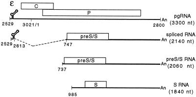
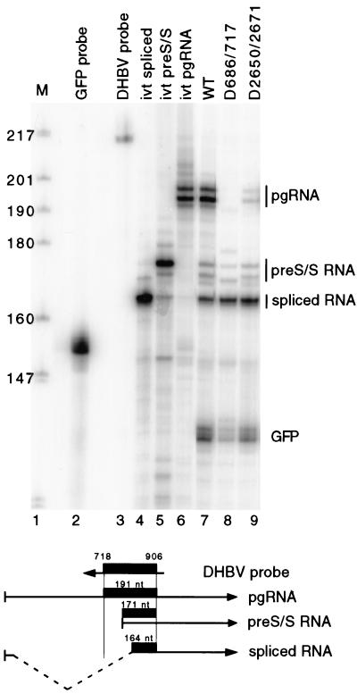
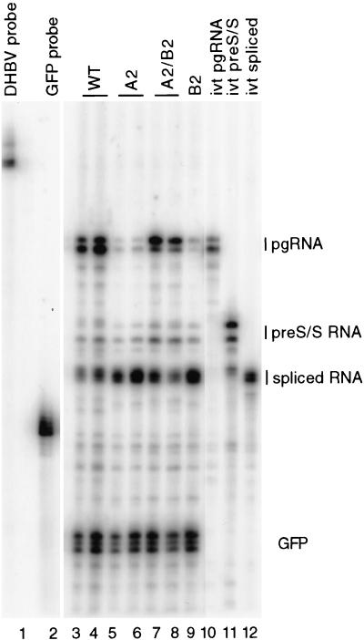
Pregenomic RNA (pgRNA) plays two major roles in the hepadnavirus life cycle. It is the mRNA for two proteins required for DNA replication, C and P, and it is the template for reverse transcription. pgRNA is a terminally redundant transcript whose synthesis does not involve RNA splicing. For duck hepatitis B virus (DHBV), a spliced RNA is derived from pgRNA by removal of a single intron. The mechanism for the simultaneous cytoplasmic accumulation of unspliced (pgRNA) and spliced RNA was not known. We found that mutations within two regions of the DHBV genome reduced the level of pgRNA while increasing the level of spliced RNA. One region is near the 5' end of pgRNA (region A), while the second is near the middle of pgRNA (region B). Inspection of the DHBV nucleotide sequence indicated that region A could base pair with region B. The 5' and 3' splice sites of the intron of the spliced RNA are within regions A and B, respectively. Substitutions that disrupted the predicted base pairing reduced the accumulation of pgRNA and increased the accumulation of spliced RNA. Restoration of base pairing, albeit mutant in sequence, resulted in restoration of pgRNA accumulation with a decrease in the level of spliced RNA. Our data are consistent with a model in which splicing of the pgRNA is suppressed by a secondary structure between regions A and B that occludes the splicing machinery from modifying pgRNA.


Figures

FIG. 1.
FIG. 1.
RNA transcription map of DHBV representing pgRNA and the three subgenomic transcripts. Coordinates of termini and locations of key features are indicated. The size of each RNA is indicated below its name. Indicated on pgRNA is the packaging signal, epsilon, and C and P genes. The initiation codons for the C and P genes are at nt 2547 and 170, respectively, and the termination codons for the C and P genes are at nt 412 and 2528, respectively. Epsilon is from nt 2560 to 2616. Indicated on spliced RNA is the position of the intron and the pre-S/S gene, which codes for the L protein. The start codons for the L and S proteins are at nt 800 and 1284, respectively, while their common termination codon is at nt 1785.
FIG. 2.
FIG. 2.
Northern blotting indicates that deletion variants do not accumulate pgRNA. Deletion of nt 2650 to 2671 or nt 686 to 717 results in accumulation of less pgRNA. Extending the nt 686-to-717 deletion to nt 840 restores accumulation of pgRNA. LMH cells were transfected with plasmids to express DHBV and GFP RNA, respectively. Poly(A)+ RNA was isolated from cells and analyzed by Northern blotting. Lane 1, wild-type RNA standard; lane 2, D686-717 RNA; lane 3, D2650-2671 RNA; lane 4, D686-840 RNA. Blots were hybridized with two probes, one that detects DHBV plus-strand sequence between nt 988 and 1665 and one that detects GFP mRNA. Positions of pregenomic class (PG), subgenomic class (SG), and GFP RNA are indicated on the left side.
FIG. 3.
FIG. 3.
RNase protection analysis of deletion variants indicates that reduction of pgRNA is due to splicing. (Top) Autoradiogram of RNase protection analysis. Lane 1, end-labeled DNA markers, MspI digest of pBR322 plasmid DNA. Sizes are indicated on the left. Lane 2, full-length GFP probe; lane 3, full-length DHBV probe; lanes 4 to 9, samples analyzed by RNase protection. Lane 4, in vitro-transcribed spliced RNA; lane 5, in vitro-transcribed pre-S/S RNA; lane 6, in vitro-transcribed pgRNA; lane 7, wild-type DHBV RNA from transfected LMH cells; lane 8, D686/717 RNA from transfected LMH cells; lane 9, D2650/2671 RNA from transfected LMH cells. Positions of protected fragments from pgRNA, pre-S/S RNA, spliced RNA, and GFP RNA are indicated on the right. (Bottom) Representation of antisense DHBV probe and the three different DHBV RNAs. The thick black line represents the probe fragment protected after RNase digestion.
FIG. 4.
FIG. 4.
Predicted secondary structure of pgRNA. (A) Predicted base pairing between regions A and B. Nucleotide coordinates are indicated. Splice donor and acceptor sites are indicated with arrows. Extent of deletion in D2650/2671 and D686/717 clones is shown. (B) Location of regions A and B on pgRNA. Region A is 75 nt from the 5′ end of pgRNA. Region B is 1,030 nt 3′ of region A.
FIG. 5.
FIG. 5.
Disruption of base pairing between regions A and B results in accumulation of lower levels of pgRNA; restoration of base pairing increases the accumulation of pgRNA. (A) The A2 and B2 variants have 6 nt substituted. (B) Northern blotting of 2-series variants. Poly(A)+ RNA from transfected LMH cells was analyzed. Lane 1, wild-type standard; lane 2, B2 variant; lane 3, A2/B2 variant; lane 4, A2 variant. Positions of pgRNA (PG), subgenomic RNA (SG), and GFP RNA are indicated on the left. The blot was hybridized with plus-strand-specific probes that detect DHBV nt 988 to 1665 and GFP mRNA.
FIG. 6.
FIG. 6.
The A/B secondary structure suppresses splicing to positively affect accumulation of pgRNA. Autoradiogram results of an RNase protection assay of the 2-series variants are shown. Poly(A)+ RNA was isolated from transfected LMH cells and analyzed. Lane 1, full-length DHBV probe; lane 2, full-length GFP probe; lanes 3 and 4, wild-type DHBV RNA; lanes 5 and 6, independently isolated molecular clones of A2 variant; lanes 7 and 8, independently isolated molecular clones of A2/B2 variant; lane 9, B2 variant; lane 10, in vitro-transcribed pgRNA; lane 11, in vitro-transcribed pre-S/S RNA; lane 12, in vitro-transcribed spliced RNA. Positions of protected fragments from pgRNA, pre-S/S RNA, spliced RNA, and GFP RNA are indicated on the right.
FIG. 7.
FIG. 7.
Northern blot analysis of 3-, 4-, and 6-series variants. Poly(A)+ RNA from transfected LMH cells was analyzed. (A) Sequence of the individual variants of the 3-, 4-, and 6-series. (B) Northern blot of 3-series variants. Lane 1, wild type; lane 2, B3 variant; lane 3, A3/B3 variant; lane 4, A3 variant. (C) Northern blot of 4-series variants. Lane 1, wild type; lane 2, B4 variant; lane 3, A4/B4 variant; lane 4, A4 variant. (D) Northern blot of 6-series variants. Lane 1, wild type; lane 2, A6 variant; lane 3, A6/B6 variant; lane 4, B6 variant. Positions of pgRNA (PG), subgenomic RNA (SG), and GFP RNA are indicated on the left. The blot was hybridized with plus-strand-specific probes that detect DHBV nt 988 to 1665 and GFP mRNA.

Similar articles

Cited by

References

    1. Beckel-Mitchener, A., and J. Summers. 1997. A novel transcriptional element in circular DNA monomers of the duck hepatitis B virus. J. Virol. 71:7917-7922. - PMC - PubMed
    1. Blanchette, M., and B. Chabot. 1997. A highly stable duplex structure sequesters the 5′ splice site region of hnRNP A1 alternative exon 7B. RNA 4:405-419. - PMC - PubMed
    1. Boris-Lawrie, K., T. M. Roberts, and S. Hull. 2001. Retroviral RNA elements integrate components of post-transcriptional gene expression. Life Sci. 69:2697-2709. - PubMed
    1. Buscher, M., W. Reiser, H. Will, and H. Schaller. 1985. Transcripts and the putative RNA pregenome of duck hepatitis B virus: implications for reverse transcription. Cell 40:717-724. - PubMed
    1. Chang, C., R. C. Hirsch, and D. Ganem. 1995. Sequences in the preC region of duck hepatitis B virus affect pregenomic RNA accumulation. Virology 207:549-554. - PubMed

Publication types

LinkOut - more resources