. 2002 Oct;76(20):10307-19.
doi: 10.1128/jvi.76.20.10307-10319.2002.

The NS2 proteins of parvovirus minute virus of mice are required for efficient nuclear egress of progeny virions in mouse cells

Affiliations

The NS2 proteins of parvovirus minute virus of mice are required for efficient nuclear egress of progeny virions in mouse cells

Virginie Eichwald et al. J Virol. 2002 Oct.

Abstract

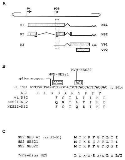
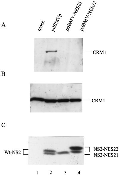
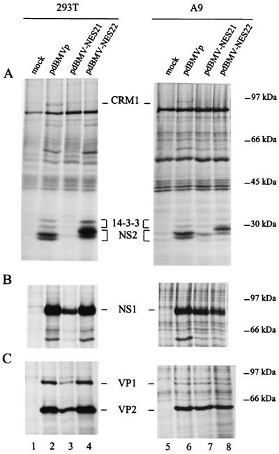
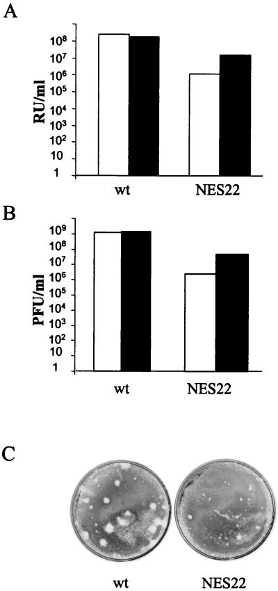
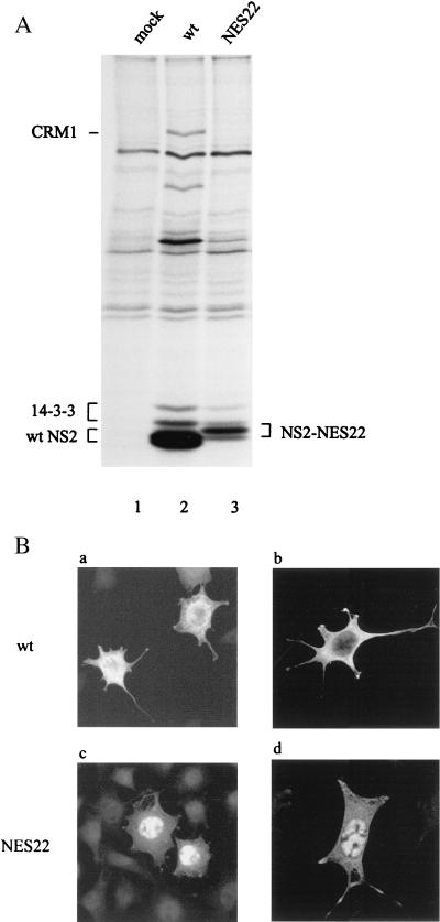
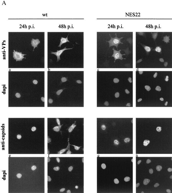
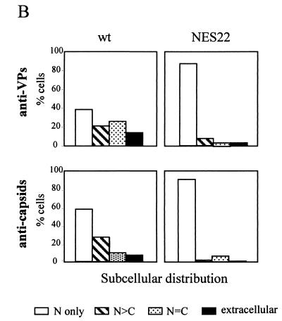
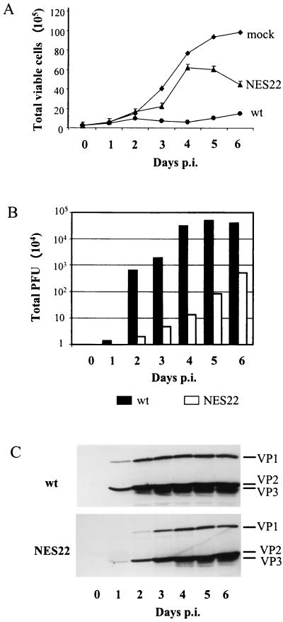
The small nonstructural NS2 proteins of parvovirus minute virus of mice (MVMp) were previously shown to interact with the nuclear export receptor Crm1. We report here the analysis of two MVM mutant genomic clones generating NS2 proteins that are unable to interact with Crm1 as a result of amino acid substitutions within their nuclear export signal (NES) sequences. Upon transfection of human and mouse cells, the MVM-NES21 and MVM-NES22 mutant genomic clones were proficient in synthesis of the four virus-encoded proteins. While the MVM-NES22 clone was further able to produce infectious mutant virions, no virus could be recovered from cells transfected with the MVM-NES21 clone. Whereas the defect of MVM-NES21 appeared to be complex, the phenotype of MVM-NES22 could be traced back to a novel distinct NS2 function. Infection of mouse cells with the MVM-NES22 mutant led to stronger nuclear retention not only of the NS2 proteins but also of infectious progeny MVM particles. This nuclear sequestration correlated with a severe delay in the release of mutant virions in the medium and with prolonged survival of the infected cell populations compared with wild-type virus-treated cultures. This defect could explain, at least in part, the small size of the plaques generated by the MVM-NES22 mutant when assayed on mouse indicator cells. Altogether, our data indicate that the interaction of MVMp NS2 proteins with the nuclear export receptor Crm1 plays a critical role at a late stage of the parvovirus life cycle involved in release of progeny viruses.


Figures

FIG. 1.
FIG. 1.
Mutagenesis strategy in the NES site of NS2. (A) The line scheme of the single-stranded MVMp genome shows the positions of the nonstructural gene promoter (P4) and the capsid gene promoter (P38). The three classes of RNA transcripts encoded by MVM, denoted R1 through R3, with introns (∧), polyadenylation sites (AAA), and open reading frames (boxes) encoding NS1, NS2, VP1, and VP2 proteins, are aligned below. The two black bars below the R2 transcript indicate the position of the NES site within the NS2 coding sequence. The stippled frame indicates the region that is shown in greater detail in panel B. (B) DNA sequence between MVMp nt 1981 and 2014 and amino acid sequence for this region of open reading frames 3 (for NS1) and 2 (for NS2). NS2 exon 2 initiates at the indicated large intron splice acceptor site. Mutations introduced into this sequence to create the two MVM-NES(−) genomes are shown in boxes above the wild-type sequence, and the resulting substitutions within the NS2 coding sequence are indicated in the last two lines, with the modified amino acids highlighted in bold. (C) Alignment of the wild-type and mutated NS2 NES sites with the NES consensus sequence (9). Leucine-like hydrophobic residues are shown in bold, and amino acid substitutions within the mutated NS2 NES sequences are shown in italics. x, any amino acid; Z, Met, Leu, Ile, Phe, or Val.
FIG. 2.
FIG. 2.
The NES21 and NES22 substitutions within the NS2 NES site prevent the interaction of NS2 with Crm1 in vivo. 293T cells were transfected with pdBMVp, pdBMV-NES21, pdBMV-NES22, or no DNA (mock) and further incubated for 48 h. (A) Whole-cell lysates were first immunoprecipitated with an NS2-specific antiserum and then immunoblotted with anti-Crm1 antibodies. As controls, nonimmunoprecipitated lysates (10% of the amount used for panel A) were analyzed by immunoblotting with either the anti-Crm1 (B) or anti-NS2 (C) antibodies. Proteins were separated on SDS-containing bipartite 8 and 12% polyacrylamide gels. Immunoblots were visualized by chemiluminescence.
FIG. 3.
FIG. 3.
Effect of the NES21 and NES22 substitutions within the NS2 NES site on viral protein synthesis. Lysates of 35S-labeled human 293T and mouse A9 cells, transfected with pdBMVp, pdBMV-NES21, pdBMV-NES22, or no DNA (mock), were precipitated with antisera directed against the NS2 C terminus (A), the NS1 C terminus (B), or the VP1 and VP2 C termini (C). The autoradiograms show immunoprecipitated proteins after separation by electrophoresis through SDS-containing bipartite 8 and 12% polyacrylamide gels. The positions of Crm1 (110 kDa), 14-3-3 (30 and 32 kDa), NS2 (ca. 25 kDa), NS1 (83 kDa), VP1 (83 kDa), and VP2 (65 kDa) proteins are indicated. Molecular masses of prestained standard proteins are shown on the right.
FIG. 4.
FIG. 4.
The MVM-NES22 genomic clone generates the production of infectious virions in human 293T and mouse A9 cells. Cells were transfected with either pdBMVp or pdBMV-NES22 plasmid DNA and viruses were harvested in the same final volume 6 days after transfection. Virus stocks were used to infect A9 indicator cells, and virus yields were determined by performing either hybridization assays, for which titers are given in RU per milliliter of crude extract (A), or plaque assays, for which titers are given in PFU per milliliter of crude extract (B). White bars, virus stocks generated in A9 cells; black bars, virus stocks generated in 293T cells. (C) Typical size appearance of plaques obtained upon infection of A9 indicator cells with viruses generated in A9 cells. Similar patterns were observed upon infection of A9 indicator cells with viruses generated in 293T cells (data not shown). wt, wild-type MVMp; NES22, MVM-NES22 mutant.
FIG. 5.
FIG. 5.
NS2 proteins expressed from the MVM-NES22 mutant exhibit an NES(−) phenotype. (A) Coimmunoprecipitation assays from infected mouse cell extracts. Lysates of 35S-labeled A9 cells infected with wild-type MVMp (MOI = 10 RU/cell), MVM-NES22 mutant (MOI = 30 RU/cell), or no virus were immunoprecipitated with an NS2-specific antiserum. The autoradiogram shows immunoprecipitated proteins after separation by electrophoresis through an SDS-containing bipartite 8 and 12% polyacrylamide gel. (B) Subcellular distribution of wild-type and NES22-modified NS2 proteins. A9 cells were infected as described above and further analyzed by indirect immunofluorescence assay 24 h after infection, using an NS2-specific antiserum. Cells were examined by either epifluorescence (a and c) or confocal (b and d) microscopy. mock, no virus; wt, wild-type MVMp; NES22, MVM-NES22 mutant.
FIG. 6.
FIG. 6.
MVM-NES22 particles are retained in the nucleus of infected mouse cells. (A) A9 cells, infected with either wild-type MVMp (MOI = 10 RU/cell) or the MVM-NES22 mutant (MOI = 30 RU/cell), were analyzed by indirect immunofluorescence assay at 24 and 48 h postinfection, using antisera directed against the structural proteins (anti-VPs) or intact capsids (anti-capsids), and were examined by epifluorescence microscopy. The images shown are representative of the most abundant phenotypes found in the wild-type and mutant infected cells. Panels: a, VP staining in both the nucleus and cytoplasm; b and c, VP staining predominantly in the nucleus; d, VP staining in the nucleus only; e, g, and h, capsid staining in the nucleus only; f, capsid staining in both the nucleus and cytoplasm. Nuclei were visualized within each field by staining the DNA with DAPI. (B) Subcellular distribution of the structural protein (top) and capsid (bottom) antigens. Cells were treated as for panel A except for the addition of neuraminidase 4 h after virus inoculation to prevent secondary rounds of infection. The values are the percentages of at least 90 immunofluorescence-positive cells counted 48 h after infection. Scored cells were classified into four categories as follows: immunofluorescent staining that was exclusively nuclear (N), mainly nuclear (N > C), comparably nuclear and cytoplasmic (N = C), and at the periphery of infected cells (extracellular). wt, wild-type MVMp; NES22, MVM-NES22 mutant.
FIG. 6.
FIG. 6.
MVM-NES22 particles are retained in the nucleus of infected mouse cells. (A) A9 cells, infected with either wild-type MVMp (MOI = 10 RU/cell) or the MVM-NES22 mutant (MOI = 30 RU/cell), were analyzed by indirect immunofluorescence assay at 24 and 48 h postinfection, using antisera directed against the structural proteins (anti-VPs) or intact capsids (anti-capsids), and were examined by epifluorescence microscopy. The images shown are representative of the most abundant phenotypes found in the wild-type and mutant infected cells. Panels: a, VP staining in both the nucleus and cytoplasm; b and c, VP staining predominantly in the nucleus; d, VP staining in the nucleus only; e, g, and h, capsid staining in the nucleus only; f, capsid staining in both the nucleus and cytoplasm. Nuclei were visualized within each field by staining the DNA with DAPI. (B) Subcellular distribution of the structural protein (top) and capsid (bottom) antigens. Cells were treated as for panel A except for the addition of neuraminidase 4 h after virus inoculation to prevent secondary rounds of infection. The values are the percentages of at least 90 immunofluorescence-positive cells counted 48 h after infection. Scored cells were classified into four categories as follows: immunofluorescent staining that was exclusively nuclear (N), mainly nuclear (N > C), comparably nuclear and cytoplasmic (N = C), and at the periphery of infected cells (extracellular). wt, wild-type MVMp; NES22, MVM-NES22 mutant.
FIG. 7.
FIG. 7.
Delayed cell killing, virus release, and subsequent internalization in MVM-NES22 mutant-infected mouse cell cultures. (A) Cell survival kinetics of A9 cell cultures infected with wild-type MVMp (MOI = 10 RU/cell), the MVM-NES22 mutant (MOI = 30 RU/cell), or no virus. The number of viable cells in each culture was determined by multiplying the total number of cells by the percentage of living cells assayed by trypan blue exclusion. (B) Time course of virion release in infected mouse cell cultures. A9 cells were infected with wild-type MVMp or the MVM-NES22 mutant as mentioned for panel A. The amounts of infectious particles released in the medium over time were determined by plaque assays using A9 indicator cells and were expressed as total PFU after subtraction of the day 0 value. (C) Time course appearance of VP1, VP2, and VP3 proteins in infected mouse cell extracts. A9 cells were infected with either wild-type MVMp or the MVM-NES22 mutant as mentioned for panel A. Whole-cell lysates, prepared at days 1 to 6 after infection, were analyzed by immunoblotting with an antiserum directed against the common C terminus of the VP polypeptides following protein separation on SDS-containing 10% polyacrylamide gels. Immunoblots were visualized by chemiluminescence. The experiments represented in all three panels were done in parallel. mock, no virus; wt, wild-type MVMp; NES22, MVM-NES22 mutant; p.i., postinfection.

Similar articles

Cited by

References

    1. Adachi, M., M. Fukuda, and E. Nishida. 2000. Nuclear export of MAP kinase (ERK) involves a MAP kinase kinase (MEK)-dependent active transport mechanism. J. Cell Biol. 148:849-856. - PMC - PubMed
    1. Agbandje-McKenna, M., A. L. Llamas-Saiz, F. Wang, P. Tattersall, and M. G. Rossmann. 1998. Functional implications of the structure of the murine parvovirus minute virus of mice. Structure 6:1369-1381. - PubMed
    1. Askjaer, P., A. Bachi, M. Wilm, F. R. Bischoff, D. L. Weeks, V. Ogniewski, M. Ohno, C. Niehrs, J. Kjems, I. W. Mattaj, and M. Fornerod. 1999. RanGTP-regulated interactions of CRM1 with nucleoporins and a shuttling DEAD-box helicase. Mol. Cell. Biol. 19:6276-6285. - PMC - PubMed
    1. Askjaer, P., T. H. Jensen, J. Nilsson, L. Englmeier, and J. Kjems. 1998. The specificity of the CRM1-Rev nuclear export signal interaction is mediated by RanGTP. J. Biol. Chem. 273:33414-33422. - PubMed
    1. Avalosse, B., F. Dupont, and A. Burny. 1995. Gene therapy for cancer. Curr. Opin. Oncol. 7:94-100. - PubMed

Publication types

Substances

LinkOut - more resources