. 2002 Oct;76(20):10374-82.
doi: 10.1128/jvi.76.20.10374-10382.2002.

Human T-cell lymphotropic virus type 1 p12(I) expression increases cytoplasmic calcium to enhance the activation of nuclear factor of activated T cells

Affiliations

Human T-cell lymphotropic virus type 1 p12(I) expression increases cytoplasmic calcium to enhance the activation of nuclear factor of activated T cells

Wei Ding et al. J Virol. 2002 Oct.

Abstract

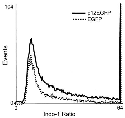
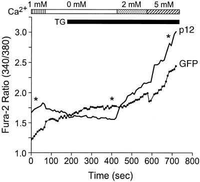
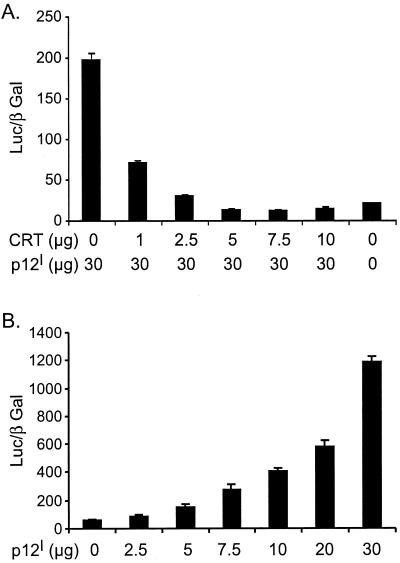
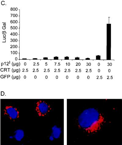
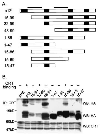
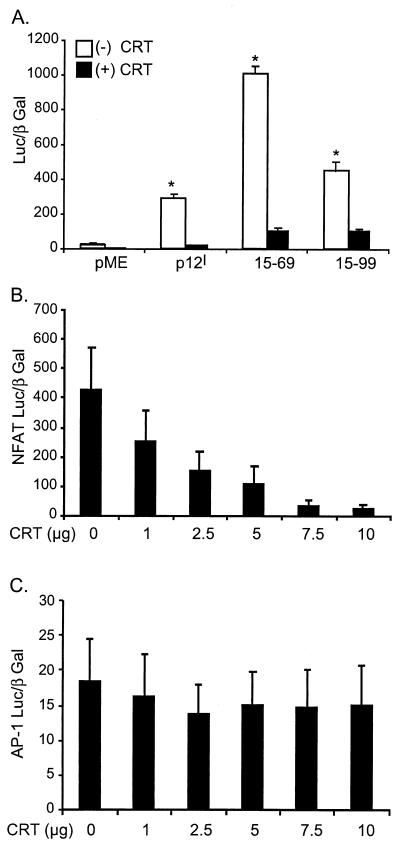
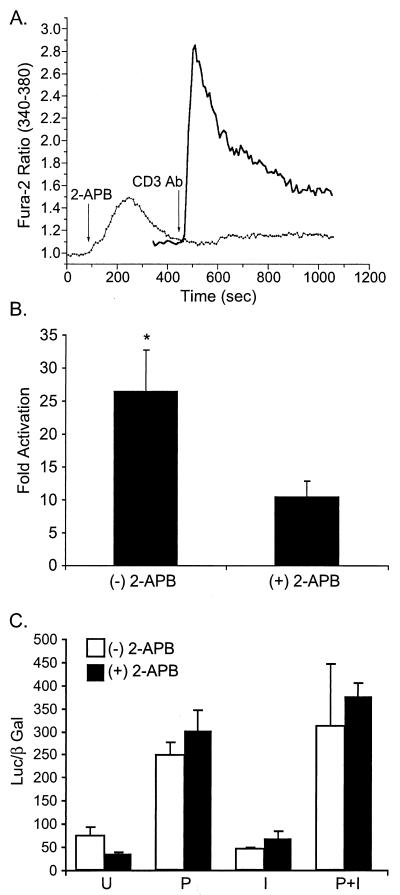
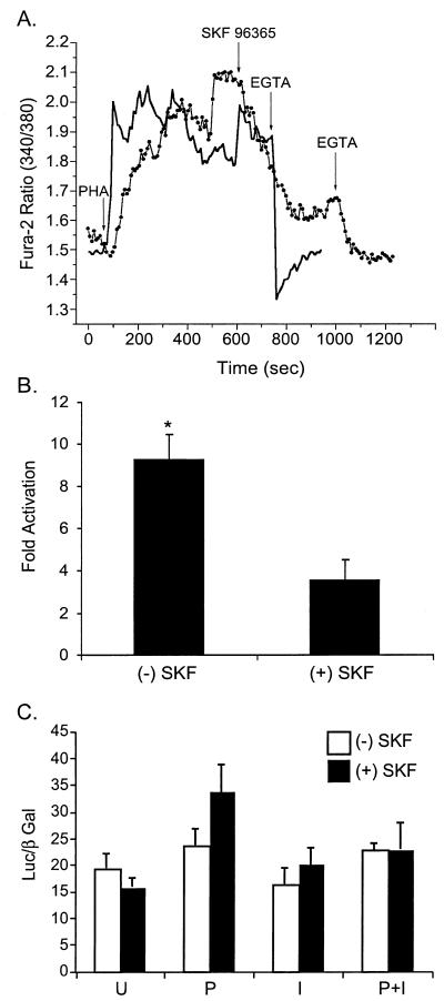
Human T-cell lymphotropic virus type 1 (HTLV-1) establishes persistent infection and is associated with lymphoproliferative or neurodegenerative diseases. As a complex retrovirus, HTLV-1 contains typical structural and enzymatic genes, as well as regulatory and accessory genes encoded in the pX region. The early events necessary for HTLV-1 to establish infection in lymphocytes, its primary target cells, remain unresolved. Recent studies have demonstrated the importance of regulatory and accessory gene products in determining this virus-host interaction. Among these, pX open reading frame I, which encodes two proteins, p12(I) and p27(I), is required for establishing persistent infection in vivo and for infection in quiescent primary lymphocytes. In addition, p12(I) localizes in the endoplasmic reticulum (ER) and cis-Golgi apparatus and associates with a calcium binding protein, calreticulin. We recently reported that p12(I) expression induces the calcium-responsive T-cell transcription factor, nuclear factor of activated T cells (NFAT), in the presence of phorbol ester activation. Based on these studies, we hypothesize that p12(I) may modulate calcium release from the ER. Here, we report that p12(I) expression increases basal cytoplasmic calcium and concurrently diminishes calcium available for release from the ER stores. Overexpression of calreticulin, a calcium buffer protein, blocked p12(I)-mediated NFAT activation independently of its ability to bind p12(I). Chemical inhibition studies using inhibitors of inositol 1,4,5-triphosphate receptor and calcium release-activated calcium channels suggest that inositol 1,4,5-triphosphate receptor in the ER membrane and calcium release-activated calcium channels in the plasma membrane contribute to p12(I)-mediated NFAT activation. Collectively, our results are the first to demonstrate the role of p12(I) in elevating cytoplasmic calcium, an antecedent to T-cell activation, and further support the important role of this accessory protein in the early events of HTLV-1 infection.


Figures

FIG. 1.
FIG. 1.
p12I increases the basal intracellular calcium in transfected Jurkat T cells. Jurkat T cells transiently transfected with p12EGFP or pEGFP-N1 plasmid were stained with indo-1/AM at 24 h posttransfection and the ratio of indo-1 emissions at 405 and 495 nm on GFP-positive cells was measured by an ELITE ESP flow cytometer. The mean ratio (10.27 ± 0.23; mean ± SEM; n = 3) in p12EGFP-transfected cells was slightly higher than the ratio in vector control pEGFP-N1-transfected cells (9.13 ± 0.58; mean ± SEM; n = 3). These data are representative of three independent experiments.
FIG. 2.
FIG. 2.
p12I diminishes calcium release from ER stores and subsequently enhances extracellular-calcium entry. Jurkat T cells transiently transfected with p12EGFP or pEGFP-N1 were loaded with fura-2/AM 48 h posttransfection, and the basal [Ca2+]i, calcium release following TG addition, and calcium entry were measured as described in Materials and Methods. The data points along the curves are means of four experiments. Statistical significances were analyzed by Student's t test. ∗, P < 0.05. Solid line, p12EGFP-transfected Jurkat T cells; dotted line, vector-transfected Jurkat T cells. Note that Jurkat T cells expressing p12EGFP have relatively higher basal [Ca2+]i and calcium entry but lower calcium release following TG addition than the control cells expressing EGFP.
FIG. 3.
FIG. 3.
Calreticulin (CRT) expression blocks p12I-mediated NFAT activation. (A) Calreticulin blocks NFAT activation induced by p12I in a dose-dependent manner. Jurkat T cells were transfected with 30 μg of pME-p12 plus increasing amounts of pcDNA-CRT plasmid, as well as NFAT reporter plasmid, and the luciferase activities were analyzed 18 h post-PMA treatment using the method described in Materials and Methods. (B) p12I dose-dependently activates NFAT in transfected Jurkat T cells. (C) Transfection with 2.5 μg of calreticulin plasmid abolishes the dose-dependent activation of NFAT induced by p12I. The GFP vector control plasmid did not affect p12I-mediated NFAT activation. The values are the means (plus SEM) of triplicate samples of at least duplicate experiments. (D) Calreticulin expression in HeLa-Tat cells is in the perinuclear region (red), consistent with its known ER localization property (27).
FIG. 3.
FIG. 3.
Calreticulin (CRT) expression blocks p12I-mediated NFAT activation. (A) Calreticulin blocks NFAT activation induced by p12I in a dose-dependent manner. Jurkat T cells were transfected with 30 μg of pME-p12 plus increasing amounts of pcDNA-CRT plasmid, as well as NFAT reporter plasmid, and the luciferase activities were analyzed 18 h post-PMA treatment using the method described in Materials and Methods. (B) p12I dose-dependently activates NFAT in transfected Jurkat T cells. (C) Transfection with 2.5 μg of calreticulin plasmid abolishes the dose-dependent activation of NFAT induced by p12I. The GFP vector control plasmid did not affect p12I-mediated NFAT activation. The values are the means (plus SEM) of triplicate samples of at least duplicate experiments. (D) Calreticulin expression in HeLa-Tat cells is in the perinuclear region (red), consistent with its known ER localization property (27).
FIG. 4.
FIG. 4.
Calreticulin (CRT) binds the region from aa 69 to 86 of p12I. (A) Schematic representation of full-length p12I and a series of p12I truncation mutants. Soild bars, transmembrane regions. Solid boxes, SH3 binding domains (PXXP motif). (B) 293T cells transfected with pME-p12 and p12I truncation mutants were lysed, and the cell lysates were precipitated with polyclonal calreticulin antibody followed by immunoblot assay to map the region in p12I associated with calreticulin (top). An aliquot of cell lysates (50 μg) was used to test the expression of p12I and the truncation mutants by immunoblot assay (middle). The same blot was stripped, and the expression of calreticulin was tested by immunoblot assay (bottom). The larger band (∼60 kDa) in the bottom blot represents native calreticulin. A smaller isoform of calreticulin (∼50 kDa) was also detected.
FIG. 5.
FIG. 5.
Calreticulin inhibition of p12I-mediated NFAT activation is independent of its association with p12I. (A) Calreticulin blocks the NFAT activation mediated by both a calreticulin binding mutant, 15-99, and a non-calreticulin binding mutant, 15-69. Thirty micrograms of pME-p12, p12I 15-99, or p12I 15-69 was used to transfect Jurkat T cells in the absence [(−) CRT] or presence [(+) CRT]of 2.5 μg of pcDNA-CRT, and the NFAT luciferase activities were analyzed 18 h post-PMA stimulation to test the effect of calreticulin on the NFAT activation mediated by both mutants. Statistical significances were analyzed by Student's t test. ∗, P < 0.05. (B) Calreticulin dose-dependently inhibits the NFAT activation induced by ionomycin treatment. Jurkat T cells were transfected with increasing amounts of pcDNA-CRT plus 10 μg of NFAT-Luc, and the NFAT luciferase activities were analyzed 18 h post-ionomycin treatment. (C) Calreticulin does not affect AP-1-responsive reporter gene. Jurkat T cells were transfected with increasing amounts of pcDNA-CRT plus 10 μg of AP-1 Luc, and the luciferase activities were analyzed 18 h post-ionomycin treatment. The values (arbitrary light units) are the means (plus SEM) of triplicate samples of at least duplicate experiments.
FIG. 6.
FIG. 6.
Inhibition of IP3 receptor reduces p12I-mediated NFAT activation. (A) IP3 receptor inhibitor, 2-APB, blocks intracellular-calcium increase following the CD3 antibody stimulation of Jurkat T cells. Solid line, Jurkat T cells stimulated with the CD3 antibody; dotted line, Jurkat T cells pretreated with 100 μM 2-APB 5 min before CD3 antibody stimulation. (B) 2-APB treatment 30 min before the addition of PMA inhibits the NFAT activation induced by p12I. (C) 2-APB treatment [(+) 2-APB] did not significantly affect AP-1 Luc activity. P, PMA; I, ionomycin; U, unstimulated. The values represent the data means (plus SEM) collected from two samples of three independent experiments. Statistical significances were analyzed by Student's t test. ∗, P < 0.05.
FIG. 7.
FIG. 7.
Inhibition of calcium release-activated calcium channel partially blocks p12I-mediated NFAT activation. (A) CRAC channel inhibitor, SKF 96365, reduces the length of the plateau phase induced by PHA stimulation. Solid line, Jurkat T cells stimulated with 2 μg of PHA/ml followed by EGTA addition. Dotted line, Jurkat T cells stimulated with PHA followed by the addition of SKF 96365 and, subsequently, EGTA. (B) Inhibition of CRAC channels partially blocks the NFAT activation mediated by p12I. SKF, SKF 96365; (+), present; (−), absent. The values represent the means (plus SEM) of triplicate samples of two independent experiments. Statistical significances were analyzed by Student's t test. ∗, P < 0.05. (C) SKF 96365 treatment did not affect AP-1 Luc activities. P, PMA, I, ionomycin, U, unstimulated.

Similar articles

Cited by

References

    1. Albrecht, B., N. D. Collins, M. T. Burniston, J. W. Nisbet, L. Ratner, P. L. Green, and M. D. Lairmore. 2000. Human T-lymphotropic virus type 1 open reading frame I p12I is required for efficient viral infectivity in primary lymphocytes. J. Virol. 74:9828-9835. - PMC - PubMed
    1. Albrecht, B., C. D. D'Souza, W. Ding, S. Tridandapani, K. M. Coggeshall, and M. D. Lairmore. 2002. Activation of nuclear factor of activated T cells (NFAT) by human T-lymphotropic virus type 1 (HTLV-1) accessory protein p12. J. Virol. 76:3493-3501. - PMC - PubMed
    1. Bangham, C. R. 2000. HTLV-1 infections. J. Clin. Pathol. 53:581-586. - PMC - PubMed
    1. Bergqvist, A., and C. M. Rice. 2001. Transcriptional activation of the interleukin-2 promoter by hepatitis C virus core protein. J. Virol. 75:772-781. - PMC - PubMed
    1. Berneman, Z. N., R. B. Gartenhaus, M. S. Reitz, W. A. Blattner, A. Manns, B. Hanchard, O. Ikehara, R. C. Gallo, and M. E. Klotman. 1992. Expression of alternatively spliced human T-lymphotropic virus type 1 pX mRNA in infected cell lines and in primary uncultured cells from patients with adult T-cell leukemia/lymphoma and healthy carriers. Proc. Natl. Acad. Sci. USA 89:3005-3009. - PMC - PubMed

Publication types

MeSH terms